Deposition Date
2014-03-03
Release Date
2014-09-03
Last Version Date
2024-02-28
Entry Detail
PDB ID:
4PQP
Keywords:
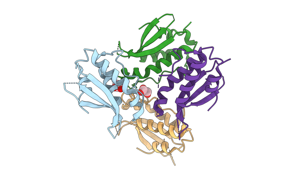
Title:
Crystal structure of human SNX14 PX domain in space group P43212
Biological Source:
Source Organism(s):
Homo sapiens (Taxon ID: 9606)
Expression System(s):
Method Details:
Experimental Method:
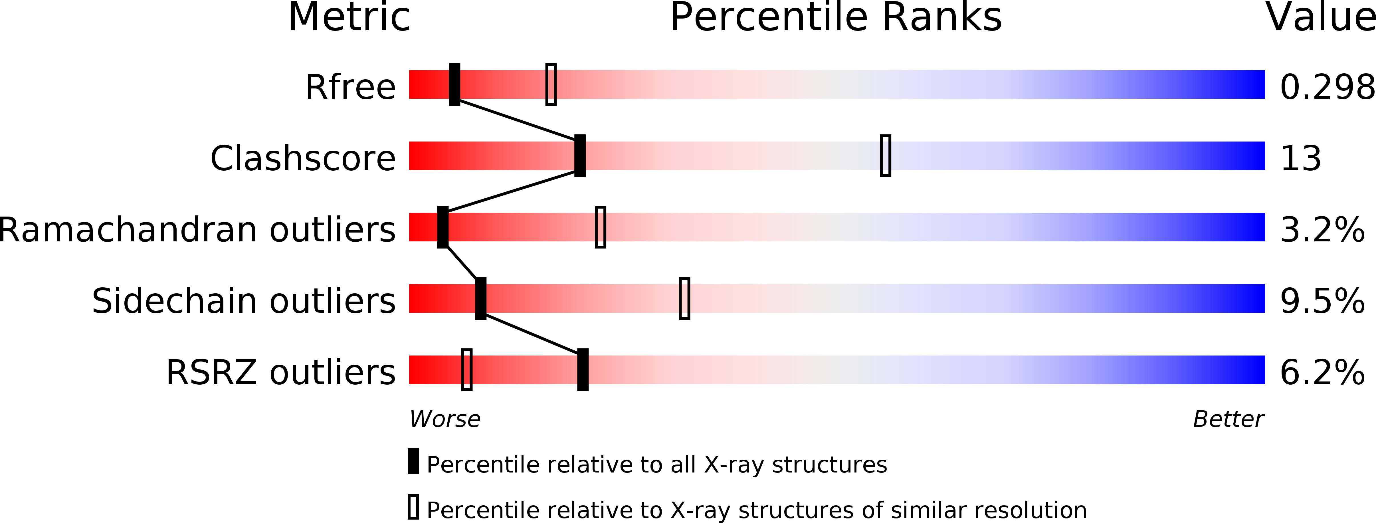
Resolution:
3.00 Å
R-Value Free:
0.28
R-Value Work:
0.23
R-Value Observed:
0.23
Space Group:
P 43 21 2