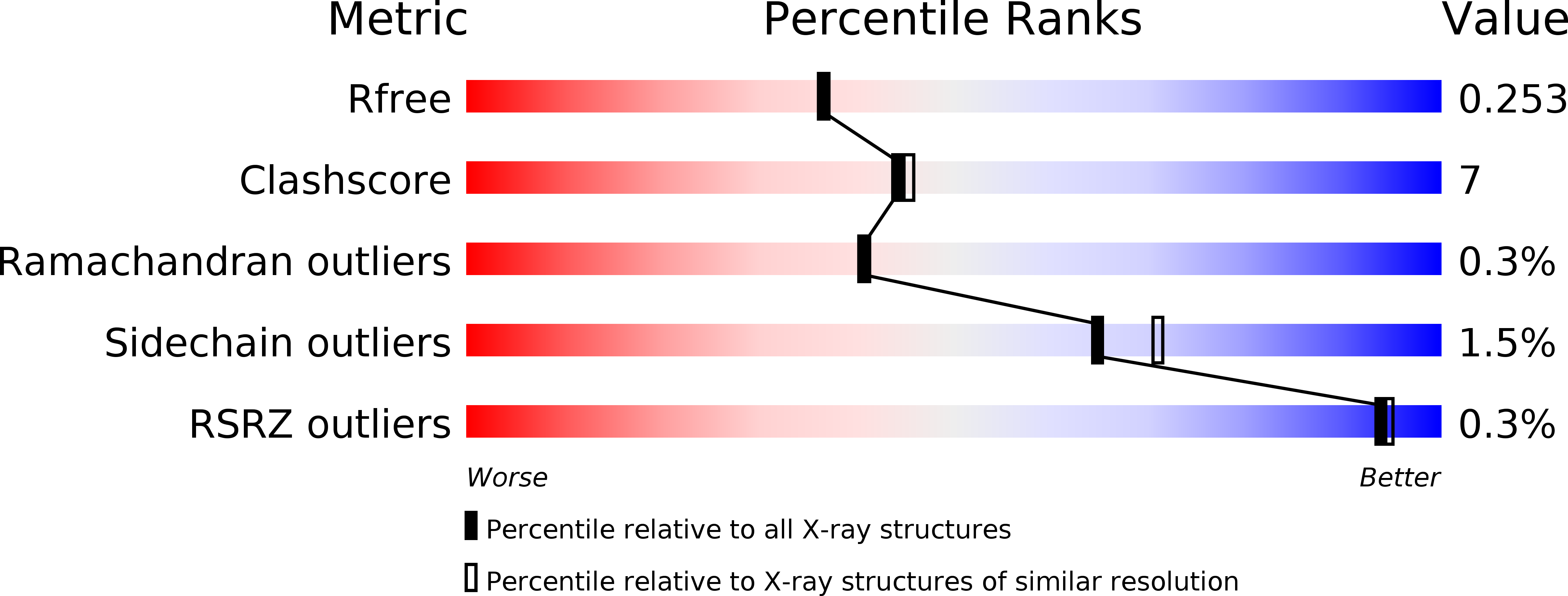
4PQ1 image
Deposition Date 2014-02-28
Release Date 2014-09-10
Last Version Date 2024-10-09
Entry Detail
PDB ID:
4PQ1
Keywords:
Title:
Crystal structure and functional implications of a DsbF homologue from Corynebacterium diphtheriae
Biological Source:
Source Organism:
Host Organism:
Method Details:
Experimental Method:
Resolution:
2.10 Å
R-Value Free:
0.24
R-Value Work:
0.18
R-Value Observed:
0.18
Space Group:
P 1 21 1
Macromolecular Entities
Polymer Type:polypeptide(L)
Molecule:Putative electron transport related protein
Gene (Uniprot):DIP0411
Chain IDs:A, B
Chain Length:158
Number of Molecules:2
Biological Source:Corynebacterium diphtheriae
Primary Citation
Structure of a DsbF homologue from Corynebacterium diphtheriae.
Acta Crystallogr.,Sect.F 70 1167 1172 (2014)
PMID: 25195886 DOI: 10.1107/S2053230X14016355

Abstact

Disulfide-bond formation, mediated by the Dsb family of proteins, is important in the correct folding of secreted or extracellular proteins in bacteria. In Gram-negative bacteria, disulfide bonds are introduced into the folding proteins in the periplasm by DsbA. DsbE from Escherichia coli has been implicated in the reduction of disulfide bonds in the maturation of cytochrome c. The Gram-positive bacterium Mycobacterium tuberculosis encodes DsbE and its homologue DsbF, the structures of which have been determined. However, the two mycobacterial proteins are able to oxidatively fold a protein in vitro, unlike DsbE from E. coli. In this study, the crystal structure of a DsbE or DsbF homologue protein from Corynebacterium diphtheriae has been determined, which revealed a thioredoxin-like domain with a typical CXXC active site. Structural comparison with M. tuberculosis DsbF would help in understanding the function of the C. diphtheriae protein.

Legend

Protein

Chemical

Disease

Primary Citation of related structures