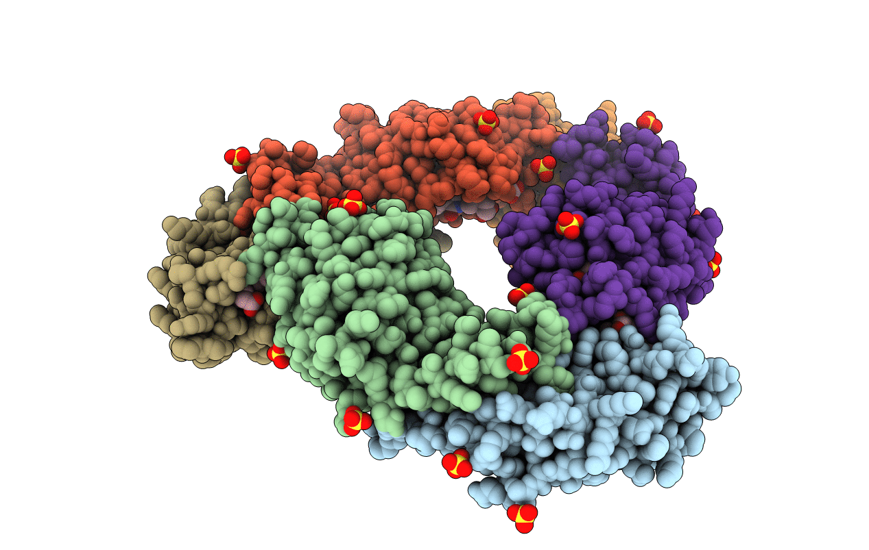
4PO5 image
Entry Detail
PDB ID:
4PO5
Keywords:
Title:
Crystal structure of allophycocyanin B from Synechocystis PCC 6803
Biological Source:
Source Organism:
PDB Version:
Deposition Date:
2014-02-24
Release Date:
2014-10-29
Method Details:
Experimental Method:
Resolution:
1.75 Å
R-Value Free:
0.19
R-Value Work:
0.17
R-Value Observed:
0.17
Space Group:
I 4 2 2
Macromolecular Entities
Polymer Type:polypeptide(L)
Description:Allophycocyanin subunit alpha-B
Chain IDs:A, C, E
Chain Length:167
Number of Molecules:3
Biological Source:Synechocystis sp.
Polymer Type:polypeptide(L)
Description:Allophycocyanin beta chain
Chain IDs:B, D, F
Chain Length:161
Number of Molecules:3
Biological Source:Synechocystis sp.
Modified Residue
Compound ID Chain ID Parent Comp ID Details 2D Image
MEN B ASN N-METHYL ASPARAGINE
Primary Citation
The structure of allophycocyanin B from Synechocystis PCC 6803 reveals the structural basis for the extreme redshift of the terminal emitter in phycobilisomes.
Acta Crystallogr.,Sect.D 70 2558 2569 (2014)
PMID: 25286841 DOI: 10.1107/S1399004714015776

Abstact

Allophycocyanin B (AP-B) is one of the two terminal emitters in phycobilisomes, the unique light-harvesting complexes of cyanobacteria and red algae. Its low excitation-energy level and the correspondingly redshifted absorption and fluorescence emission play an important role in funnelling excitation energy from the hundreds of chromophores of the extramembraneous phycobilisome to the reaction centres within the photosynthetic membrane. In the absence of crystal structures of these low-abundance terminal emitters, the molecular basis for the extreme redshift and directional energy transfer is largely unknown. Here, the crystal structure of trimeric AP-B [(ApcD/ApcB)3] from Synechocystis sp. PCC 6803 at 1.75 Å resolution is reported. In the crystal lattice, eight trimers of AP-B form a porous, spherical, 48-subunit assembly of 193 Å in diameter with an internal cavity of 1.1 × 10(6) Å(3). While the overall structure of trimeric AP-B is similar to those reported for many other phycobiliprotein trimers, the chromophore pocket of the α-subunit, ApcD, has more bulky residues that tightly pack the phycocyanobilin (PCB). Ring D of the chromophores is further stabilized by close interactions with ApcB from the adjacent monomer. The combined contributions from both subunits render the conjugated rings B, C and D of the PCB in ApcD almost perfectly coplanar. Together with mutagenesis data, it is proposed that the enhanced planarity effectively extends the conjugation system of PCB and leads to the redshifted absorption (λmax = 669 nm) and fluorescence emission (679 nm) of the ApcD chromophore in AP-B, thereby enabling highly efficient energy transfer from the phycobilisome core to the reaction centres.

Legend

Protein

Chemical

Disease

Primary Citation of related structures