4PO2 image
Deposition Date 2014-02-24
Release Date 2014-08-20
Last Version Date 2024-02-28
Entry Detail
PDB ID:
4PO2
Keywords:
Title:
Crystal Structure of the Stress-Inducible Human Heat Shock Protein HSP70 Substrate-Binding Domain in Complex with Peptide Substrate
Biological Source:
Source Organism:
Homo sapiens (Taxon ID: 9606)
(Taxon ID: )
Host Organism:
Method Details:
Experimental Method:
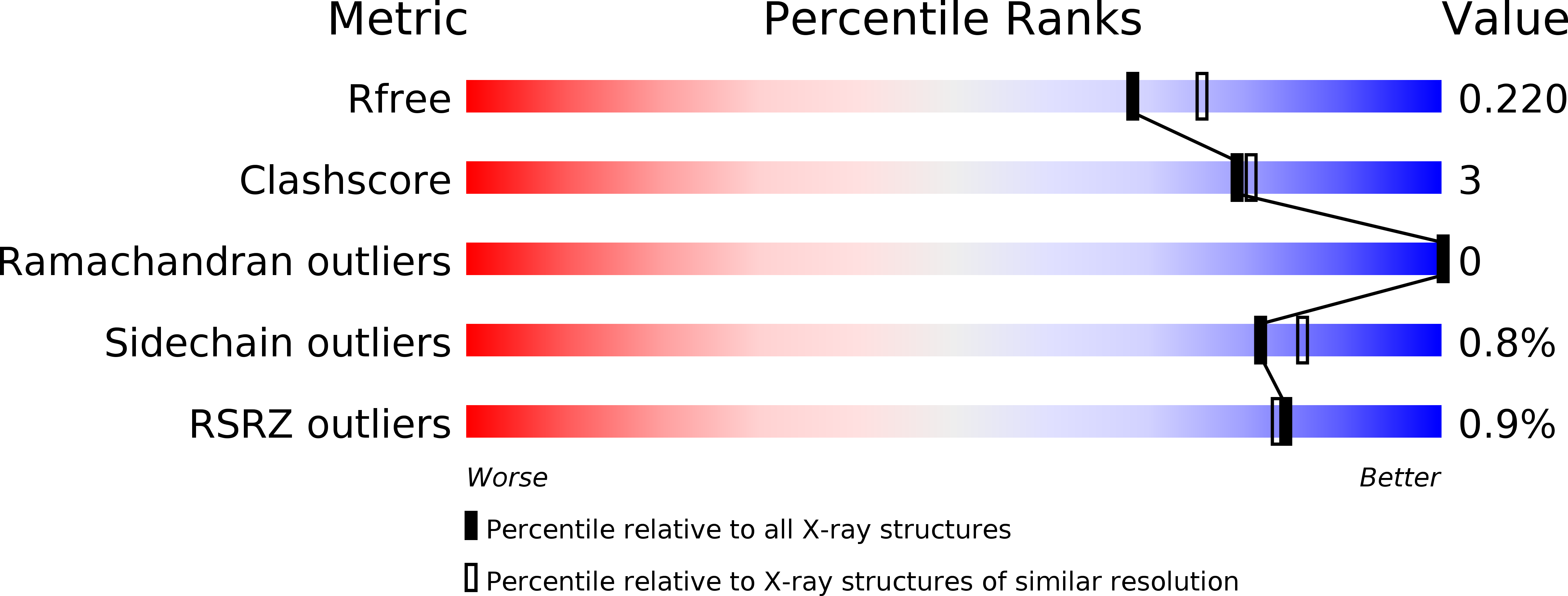
Resolution:
2.00 Å
R-Value Free:
0.22
R-Value Work:
0.17
R-Value Observed:
0.17
Space Group:
C 1 2 1
Macromolecular Entities
Polymer Type:polypeptide(L)
Molecule:Heat shock 70 kDa protein 1A/1B
Chain IDs:A, B
Chain Length:235
Number of Molecules:2
Biological Source:Homo sapiens
Polymer Type:polypeptide(L)
Molecule:HSP70 substrate peptide
Chain IDs:C, D
Chain Length:7
Number of Molecules:2
Biological Source:
Primary Citation
Crystal structure of the stress-inducible human heat shock protein 70 substrate-binding domain in complex with Peptide substrate.
Plos One 9 e103518 e103518 (2014)
PMID: 25058147 DOI: 10.1371/journal.pone.0103518

Abstact

The HSP70 family of molecular chaperones function to maintain protein quality control and homeostasis. The major stress-induced form, HSP70 (also called HSP72 or HSPA1A) is considered an important anti-cancer drug target because it is constitutively overexpressed in a number of human cancers and promotes cancer cell survival. All HSP70 family members contain two functional domains: an N-terminal nucleotide binding domain (NBD) and a C-terminal protein substrate-binding domain (SBD); the latter is subdivided into SBDα and SBDβ subdomains. The NBD and SBD structures of the bacterial ortholog, DnaK, have been characterized, but only the isolated NBD and SBDα segments of eukaryotic HSP70 proteins have been determined. Here we report the crystal structure of the substrate-bound human HSP70-SBD to 2 angstrom resolution. The overall fold of this SBD is similar to the corresponding domain in the substrate-bound DnaK structures, confirming a similar overall architecture of the orthologous bacterial and human HSP70 proteins. However, conformational differences are observed in the peptide-HSP70-SBD complex, particularly in the loop L(α, β) that bridges SBDα to SBDβ, and the loop L(L,1) that connects the SBD and NBD. The interaction between the SBDα and SBDβ subdomains and the mode of substrate recognition is also different between DnaK and HSP70. This suggests that differences may exist in how different HSP70 proteins recognize their respective substrates. The high-resolution structure of the substrate-bound-HSP70-SBD complex provides a molecular platform for the rational design of small molecule compounds that preferentially target this C-terminal domain, in order to modulate human HSP70 function.

Legend

Protein

Chemical

Disease

Primary Citation of related structures