
Deposition Date
2014-05-22
Release Date
2014-10-08
Last Version Date
2023-12-20
Entry Detail
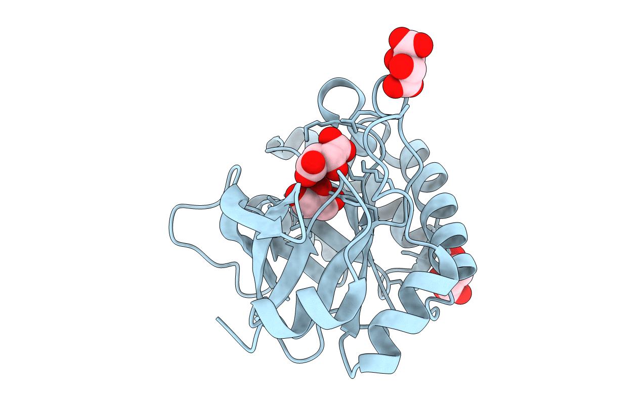
PDB ID:
4PMQ
Keywords:
Title:
Crystal structure of the Mycobacterium tuberculosis Tat-secreted protein Rv2525c in complex with L-tartrate (orthorhombic crystal form)
Biological Source:
Source Organism(s):
Mycobacterium tuberculosis (Taxon ID: 83331)
Expression System(s):
Method Details:
Experimental Method:
Resolution:
1.61 Å
R-Value Free:
0.19
R-Value Work:
0.16
R-Value Observed:
0.16
Space Group:
P 21 21 2
