4PL8 image
Entry Detail
PDB ID:
4PL8
Title:
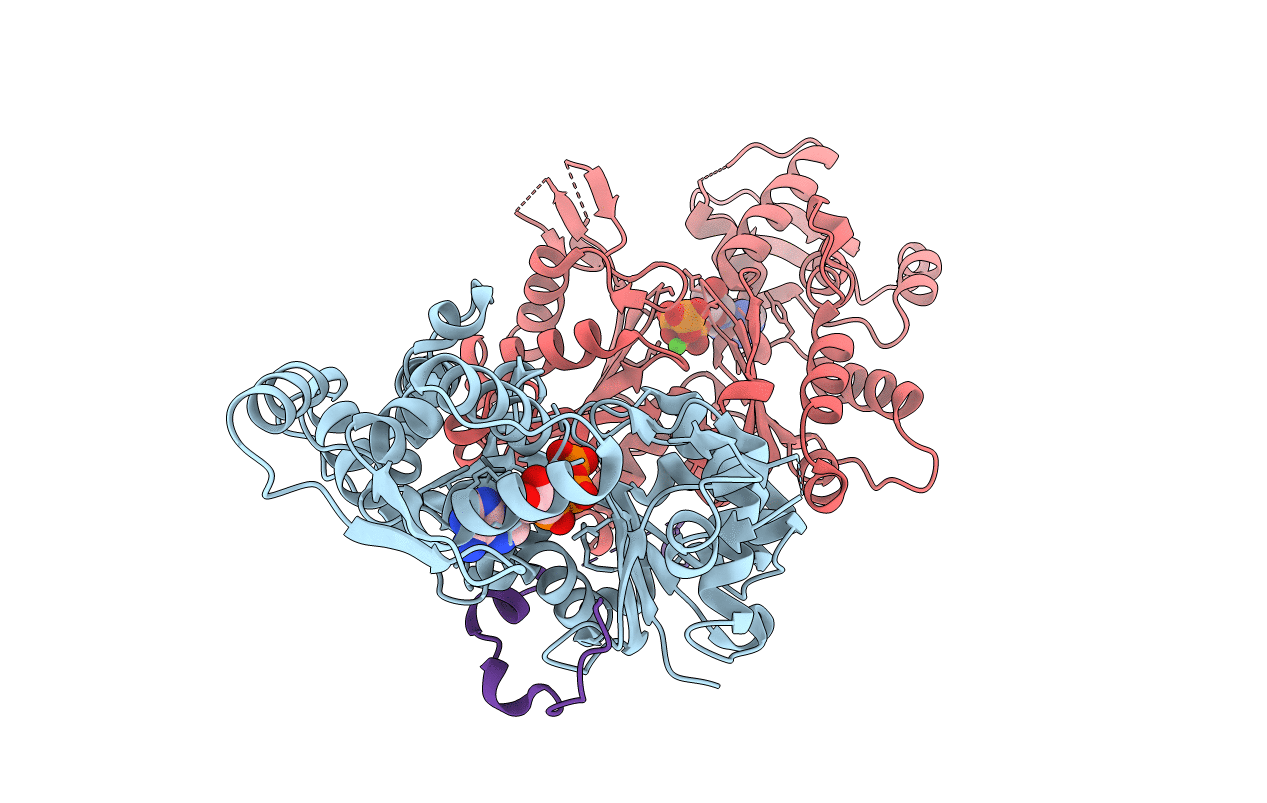
Structure of rabbit skeletal muscle actin in complex with a hybrid peptide comprising thymosin beta4 and the lysine-rich region of Cordon-Bleu
Biological Source:
PDB Version:
Deposition Date:
2014-05-16
Release Date:
2014-10-22
Method Details:
Experimental Method:
Resolution:
2.00 Å
R-Value Free:
0.20
R-Value Work:
0.17
R-Value Observed:
0.17
Space Group:
P 43 21 2
Macromolecular Entities
Polymer Type:polypeptide(L)
Description:Actin, alpha skeletal muscle
Chain IDs:A, C (auth: B)
Chain Length:375
Number of Molecules:2
Biological Source:Oryctolagus cuniculus
Polymer Type:polypeptide(L)
Description:Thymosin beta-4,Protein cordon-bleu,Thymosin beta-4
Chain IDs:B (auth: H)
Chain Length:73
Number of Molecules:1
Biological Source:Homo sapiens
Modified Residue
Compound ID Chain ID Parent Comp ID Details 2D Image
HIC A HIS modified residue
Primary Citation
Structural basis of thymosin-beta 4/profilin exchange leading to actin filament polymerization.
Proc.Natl.Acad.Sci.USA 111 E4596 E4605 (2014)
PMID: 25313062 DOI: 10.1073/pnas.1412271111

Abstact

Thymosin-β4 (Tβ4) and profilin are the two major sequestering proteins that maintain the pool of monomeric actin (G-actin) within cells of higher eukaryotes. Tβ4 prevents G-actin from joining a filament, whereas profilin:actin only supports barbed-end elongation. Here, we report two Tβ4:actin structures. The first structure shows that Tβ4 has two helices that bind at the barbed and pointed faces of G-actin, preventing the incorporation of the bound G-actin into a filament. The second structure displays a more open nucleotide binding cleft on G-actin, which is typical of profilin:actin structures, with a concomitant disruption of the Tβ4 C-terminal helix interaction. These structures, combined with biochemical assays and molecular dynamics simulations, show that the exchange of bound actin between Tβ4 and profilin involves both steric and allosteric components. The sensitivity of profilin to the conformational state of actin indicates a similar allosteric mechanism for the dissociation of profilin during filament elongation.

Legend

Protein

Chemical

Disease

Primary Citation of related structures