Deposition Date
2014-05-16
Release Date
2014-10-22
Last Version Date
2023-11-08
Entry Detail
PDB ID:
4PL7
Keywords:
Title:
Structure of Komagataella pastoris actin-thymosin beta4 hybrid
Biological Source:
Source Organism(s):
Komagataella pastoris (Taxon ID: 644223)
Homo sapiens (Taxon ID: 9606)
Homo sapiens (Taxon ID: 9606)
Expression System(s):
Method Details:
Experimental Method:
Resolution:
2.30 Å
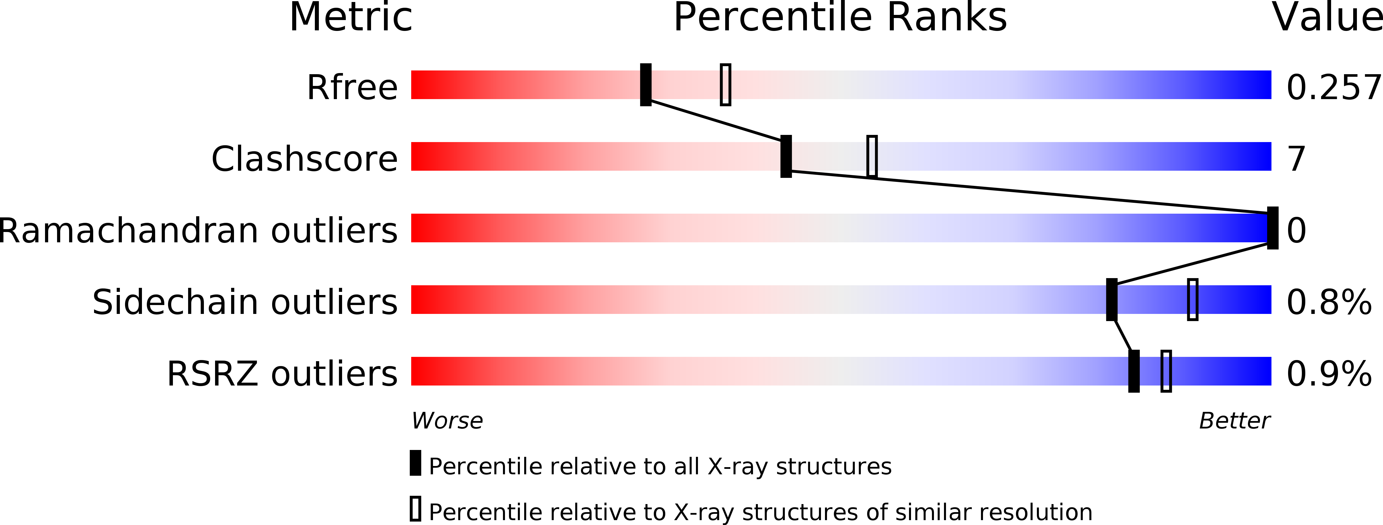
R-Value Free:
0.25
R-Value Work:
0.19
R-Value Observed:
0.19
Space Group:
P 1 21 1