4PL5 image
Entry Detail
PDB ID:
4PL5
Title:
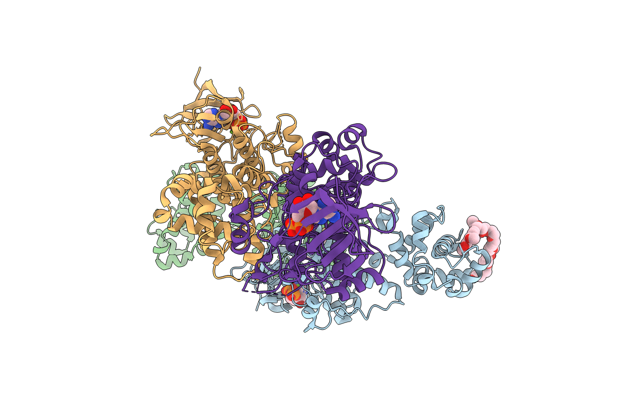
Crystal structure of murine IRE1 in complex with OICR573 inhibitor
Biological Source:
Source Organism:
Host Organism:
PDB Version:
Deposition Date:
2014-05-16
Release Date:
2014-09-03
Method Details:
Experimental Method:
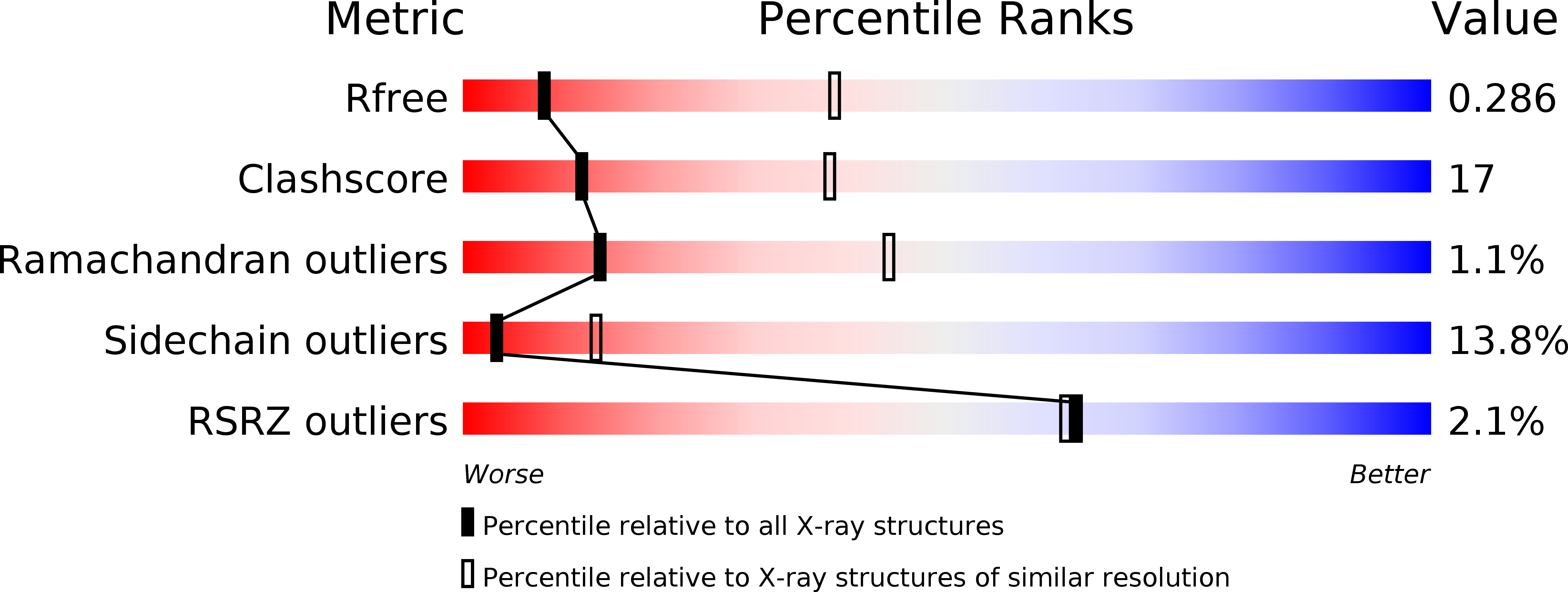
Resolution:
3.40 Å
R-Value Free:
0.28
R-Value Work:
0.22
R-Value Observed:
0.22
Space Group:
C 1 2 1
Macromolecular Entities
Polymer Type:polypeptide(L)
Description:Serine/threonine-protein kinase/endoribonuclease IRE1
Chain IDs:A (auth: B), B (auth: A), C, D
Chain Length:435
Number of Molecules:4
Biological Source:Mus musculus
Primary Citation
Structure and mechanism of action of the hydroxy-aryl-aldehyde class of IRE1 endoribonuclease inhibitors.
Nat Commun 5 4202 4202 (2014)
PMID: 25164867 DOI: 10.1038/ncomms5202

Abstact

Endoplasmic reticulum (ER) stress activates the unfolded protein response and its dysfunction is linked to multiple diseases. The stress transducer IRE1α is a transmembrane kinase endoribonuclease (RNase) that cleaves mRNA substrates to re-establish ER homeostasis. Aromatic ring systems containing hydroxy-aldehyde moieties, termed hydroxy-aryl-aldehydes (HAA), selectively inhibit IRE1α RNase and thus represent a novel chemical series for therapeutic development. We solved crystal structures of murine IRE1α in complex with three HAA inhibitors. HAA inhibitors engage a shallow pocket at the RNase-active site through pi-stacking interactions with His910 and Phe889, an essential Schiff base with Lys907 and a hydrogen bond with Tyr892. Structure-activity studies and mutational analysis of contact residues define the optimal chemical space of inhibitors and validate the inhibitor-binding site. These studies lay the foundation for understanding both the biochemical and cellular functions of IRE1α using small molecule inhibitors and suggest new avenues for inhibitor design.

Legend

Protein

Chemical

Disease

Primary Citation of related structures