Deposition Date
2014-05-15
Release Date
2014-10-08
Last Version Date
2023-12-27
Entry Detail
PDB ID:
4PKX
Keywords:
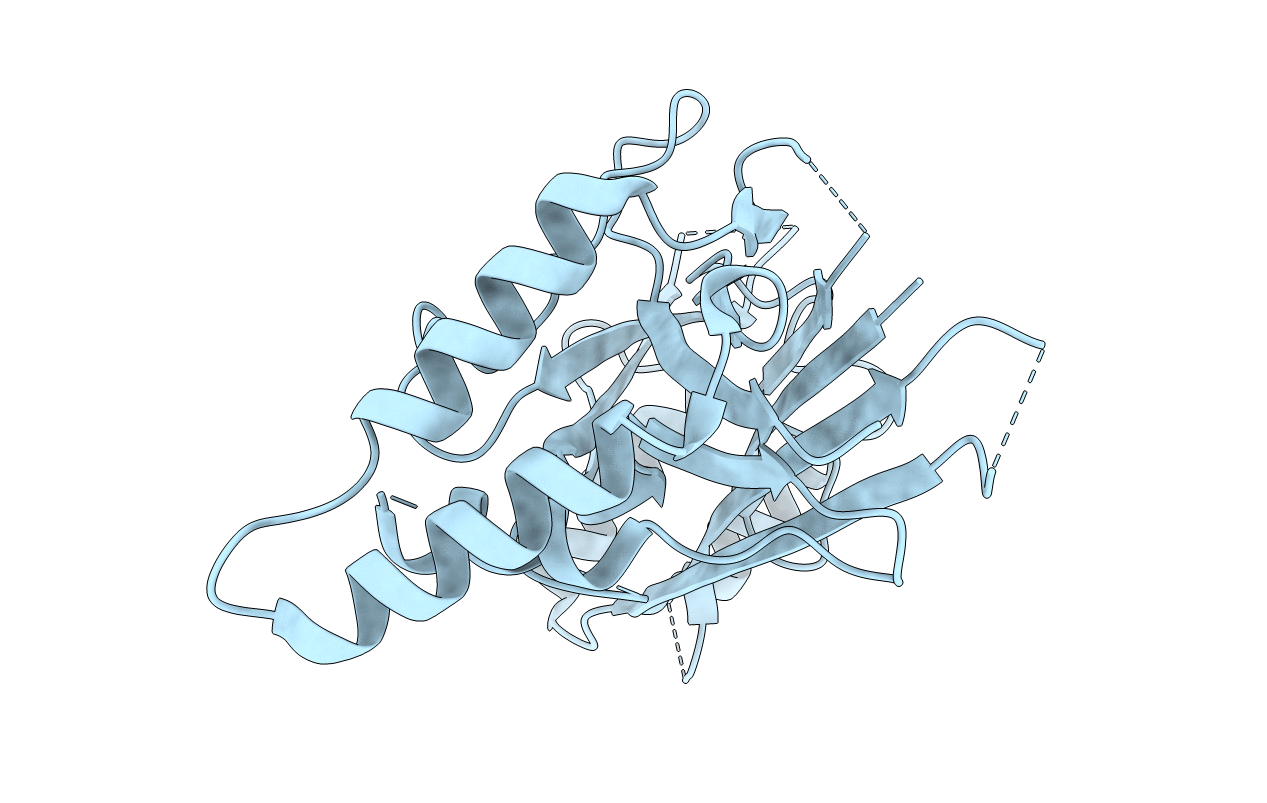
Title:
The structure of a conserved Piezo channel domain reveals a novel beta sandwich fold
Biological Source:
Source Organism(s):
Caenorhabditis elegans (Taxon ID: 6239)
Expression System(s):
Method Details:
Experimental Method:
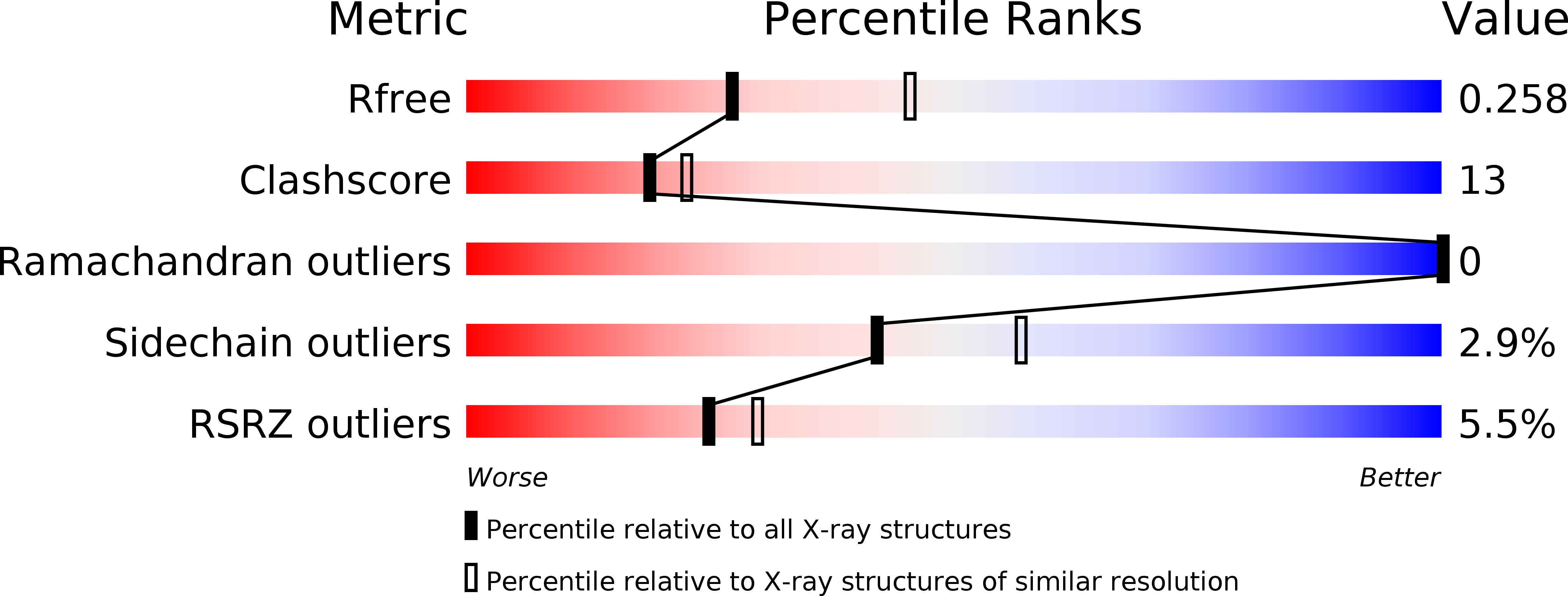
Resolution:
2.54 Å
R-Value Free:
0.25
R-Value Work:
0.21
R-Value Observed:
0.21
Space Group:
P 1 21 1