Deposition Date
2014-05-15
Release Date
2014-08-20
Last Version Date
2024-10-09
Entry Detail
PDB ID:
4PKO
Keywords:
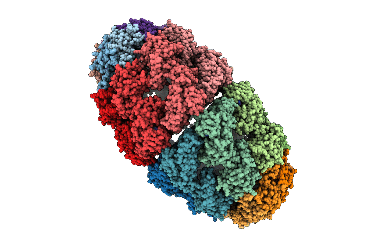
Title:
Crystal structure of the Football-shaped GroEL-GroES2-(ADPBeFx)14 complex
Biological Source:
Source Organism(s):
Escherichia coli (Taxon ID: 562)
Expression System(s):
Method Details:
Experimental Method:
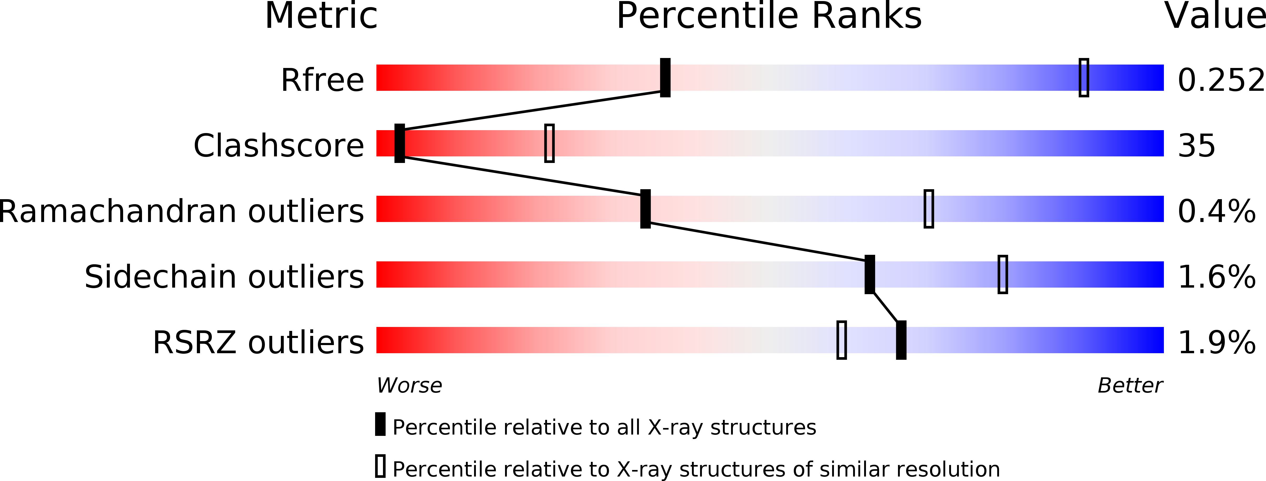
Resolution:
3.84 Å
R-Value Free:
0.24
R-Value Work:
0.18
R-Value Observed:
0.18
Space Group:
P 21 21 21