4PK6 image
Deposition Date 2014-05-13
Release Date 2014-09-03
Last Version Date 2024-11-13
Entry Detail
PDB ID:
4PK6
Title:
Crystal structure of the indoleamine 2,3-dioxygenagse 1 (IDO1) complexed with imidazothiazole derivative
Biological Source:
Source Organism:
Homo sapiens (Taxon ID: 9606)
Host Organism:
Method Details:
Experimental Method:
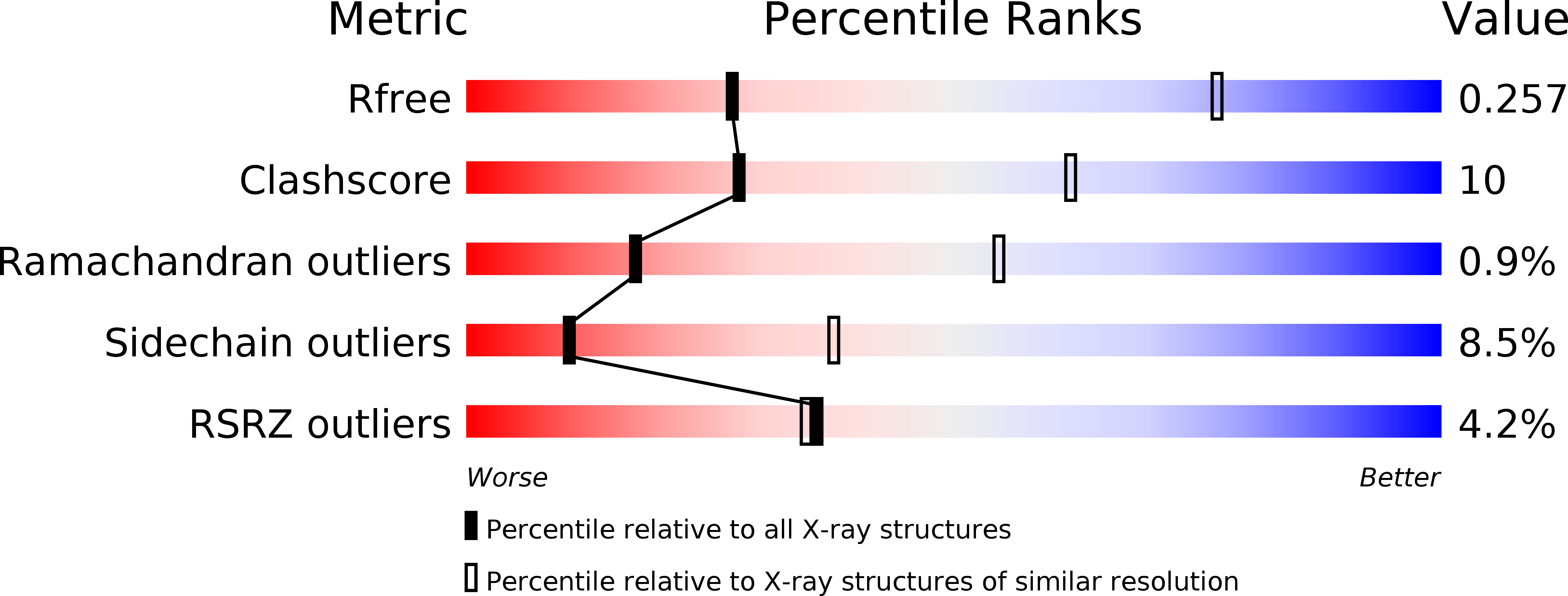
Resolution:
3.45 Å
R-Value Free:
0.25
R-Value Work:
0.18
R-Value Observed:
0.19
Space Group:
P 21 21 21
Macromolecular Entities
Structures with similar UniProt ID
Protein Blast
Polymer Type:polypeptide(L)
Molecule:Indoleamine 2,3-dioxygenase 1
Gene (Uniprot):IDO1
Chain IDs:A, B
Chain Length:423
Number of Molecules:2
Biological Source:Homo sapiens
Primary Citation
Crystal Structures and Structure-Activity Relationships of Imidazothiazole Derivatives as IDO1 Inhibitors.
Acs Med.Chem.Lett. 5 1119 1123 (2014)
PMID: 25313323 DOI: 10.1021/ml500247w

Abstact

Indoleamine 2,3-dioxygenase 1 (IDO1) is considered as a promising target for the treatment of several diseases, including neurological disorders and cancer. We report here the crystal structures of two IDO1/IDO1 inhibitor complexes, one of which shows that Amg-1 is directly bound to the heme iron of IDO1 with a clear induced fit. We also describe the identification and preliminary optimization of imidazothiazole derivatives as novel IDO1 inhibitors. Using our crystal structure information and structure-activity relationships (SAR) at the pocket-B of IDO1, we found a series of urea derivatives as potent IDO1 inhibitors and revealed that generation of an induced fit and the resulting interaction with Phe226 and Arg231 are essential for potent IDO1 inhibitory activity. The results of this study are very valuable for understanding the mechanism of IDO1 activation, which is very important for structure-based drug design (SBDD) to discover potent IDO1 inhibitors.

Legend

Protein

Chemical

Disease

Primary Citation of related structures