
Deposition Date
2014-05-09
Release Date
2014-08-06
Last Version Date
2024-10-30
Entry Detail
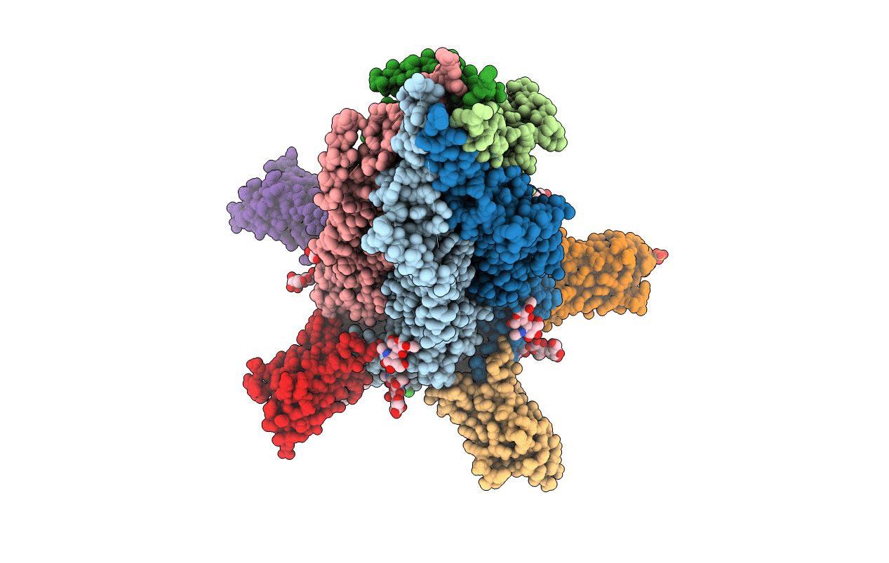
PDB ID:
4PIR
Keywords:
Title:
X-ray structure of the mouse serotonin 5-HT3 receptor
Biological Source:
Source Organism(s):
Mus musculus (Taxon ID: 10090)
Lama glama (Taxon ID: 9844)
Lama glama (Taxon ID: 9844)
Expression System(s):
Method Details:
Experimental Method:
Resolution:
3.50 Å
R-Value Free:
0.25
R-Value Work:
0.21
R-Value Observed:
0.21
Space Group:
P 21 21 21
