Deposition Date
2014-05-08
Release Date
2015-10-14
Last Version Date
2023-12-27
Entry Detail
PDB ID:
4PI9
Keywords:
Title:
Crystal structure of S. Aureus Autolysin E in complex with muropeptide NAM-L-ALA-D-iGLU
Biological Source:
Source Organism(s):
Expression System(s):
Method Details:
Experimental Method:
Resolution:
1.48 Å
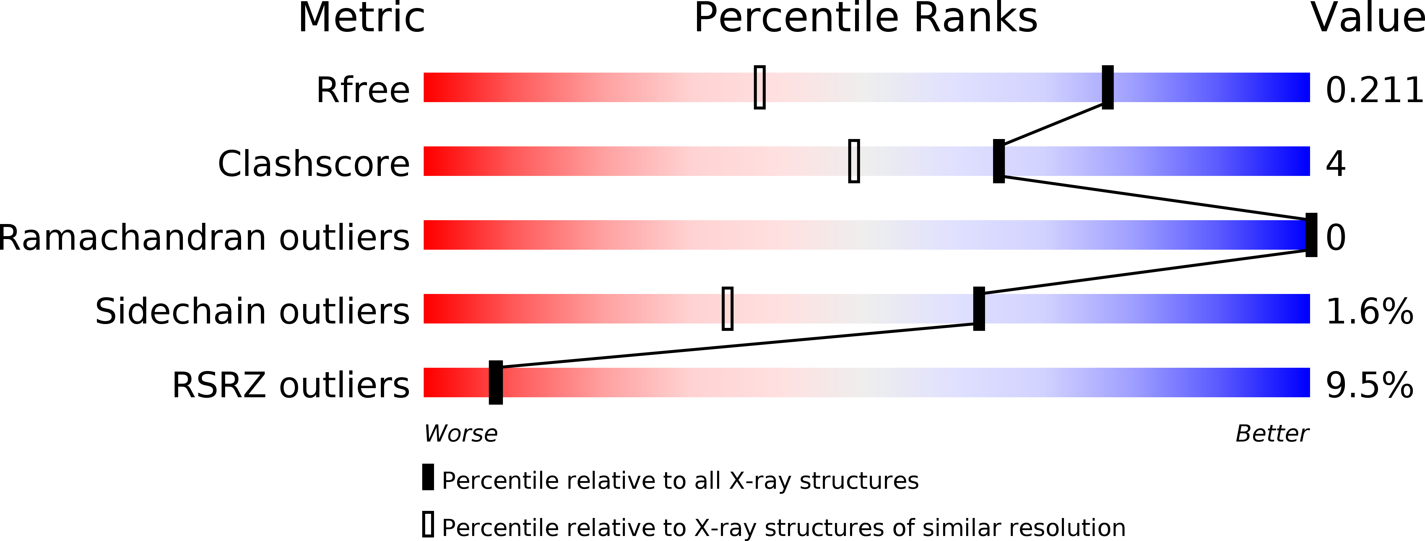
R-Value Free:
0.20
R-Value Work:
0.17
R-Value Observed:
0.17
Space Group:
P 21 21 21