4PH3 image
Deposition Date 2014-05-03
Release Date 2015-06-10
Last Version Date 2024-11-20
Entry Detail
PDB ID:
4PH3
Keywords:
Title:
N-terminal domain of the capsid protein from bovine leukaemia virus (with no beta-hairpin)
Biological Source:
Source Organism:
Host Organism:
Method Details:
Experimental Method:
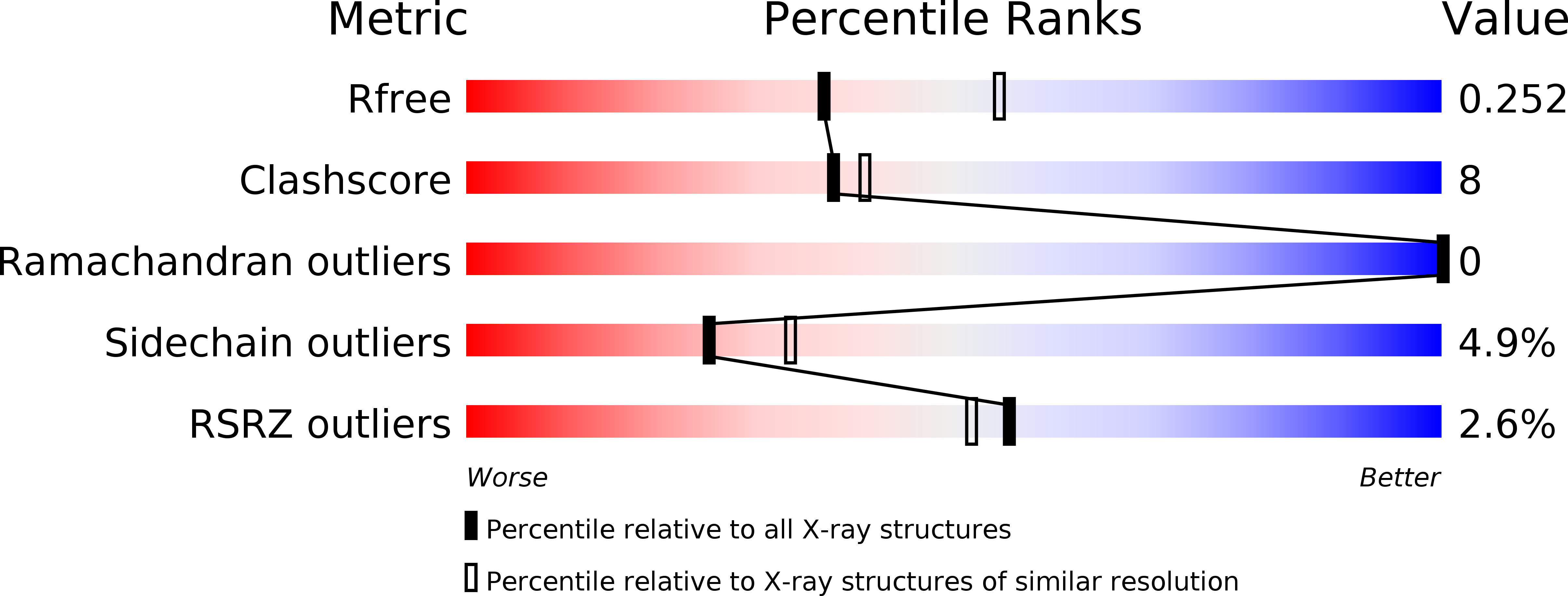
Resolution:
2.44 Å
R-Value Free:
0.24
R-Value Work:
0.20
R-Value Observed:
0.20
Space Group:
P 21 21 21
Macromolecular Entities
Structures with similar UniProt ID
Protein Blast
Polymer Type:polypeptide(L)
Molecule:BLV capsid
Gene (Uniprot):gag-pro-pol
Chain IDs:A, B
Chain Length:129
Number of Molecules:2
Biological Source:Bovine leukemia virus
Modified Residue
Compound ID Chain ID Parent Comp ID Details 2D Image
CSO A CYS modified residue
Primary Citation
STRUCTURAL VIROLOGY. Conformational plasticity of a native retroviral capsid revealed by x-ray crystallography.
Science 349 95 98 (2015)
PMID: 26044299 DOI: 10.1126/science.aaa5182

Abstact

Retroviruses depend on self-assembly of their capsid proteins (core particle) to yield infectious mature virions. Despite the essential role of the retroviral core, its high polymorphism has hindered high-resolution structural analyses. Here, we report the x-ray structure of the native capsid (CA) protein from bovine leukemia virus. CA is organized as hexamers that deviate substantially from sixfold symmetry, yet adjust to make two-dimensional pseudohexagonal arrays that mimic mature retroviral cores. Intra- and interhexameric quasi-equivalent contacts are uncovered, with flexible trimeric lateral contacts among hexamers, yet preserving very similar dimeric interfaces making the lattice. The conformation of each capsid subunit in the hexamer is therefore dictated by long-range interactions, revealing how the hexamers can also assemble into closed core particles, a relevant feature of retrovirus biology.

Legend

Protein

Chemical

Disease

Primary Citation of related structures