4PGI image
Entry Detail
PDB ID:
4PGI
Keywords:
Title:
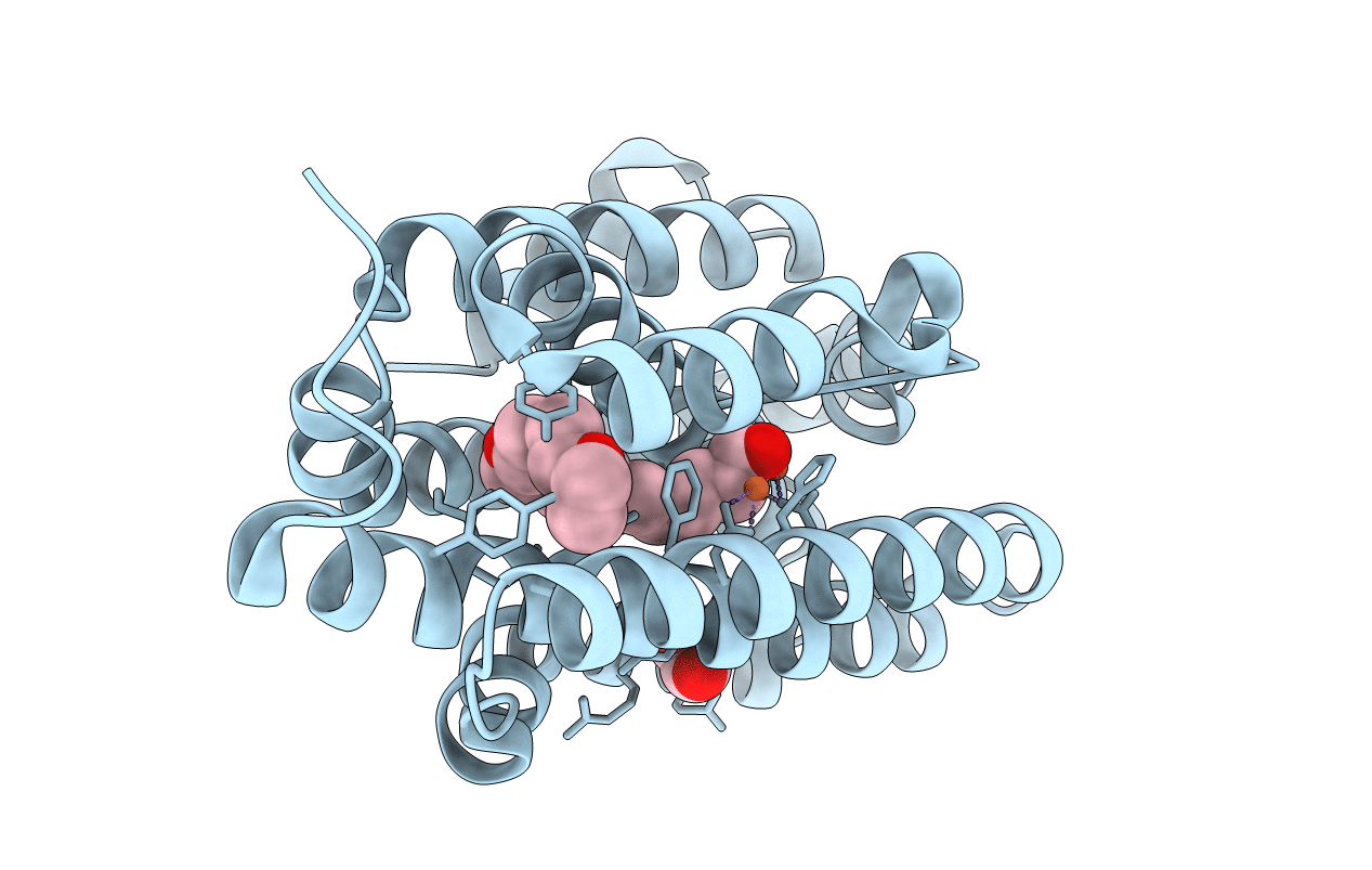
Insights into Substrate and Metal Binding from the Crystal Structure of Cyanobacterial Aldehyde Deformylating Oxygenase with Substrate Analogs Bound
Biological Source:
Source Organism:
Host Organism:
PDB Version:
Deposition Date:
2014-05-02
Release Date:
2015-02-11
Method Details:
Experimental Method:
Resolution:
2.08 Å
R-Value Free:
0.22
R-Value Work:
0.19
R-Value Observed:
0.19
Space Group:
P 43 21 2
Macromolecular Entities
Polymer Type:polypeptide(L)
Description:Aldehyde decarbonylase
Mutations:E90K, L194A
Chain IDs:A
Chain Length:250
Number of Molecules:1
Biological Source:Prochlorococcus marinus
Primary Citation
Insights into substrate and metal binding from the crystal structure of cyanobacterial aldehyde deformylating oxygenase with substrate bound.
Acs Chem.Biol. 9 2584 2593 (2014)
PMID: 25222710 DOI: 10.1021/cb500343j

Abstact

The nonheme diiron enzyme cyanobacterial aldehyde deformylating oxygenase, cADO, catalyzes the highly unusual deformylation of aliphatic aldehydes to alkanes and formate. We have determined crystal structures for the enzyme with a long-chain water-soluble aldehyde and medium-chain carboxylic acid bound to the active site. These structures delineate a hydrophobic channel that connects the solvent with the deeply buried active site and reveal a mode of substrate binding that is different from previously determined structures with long-chain fatty acids bound. The structures also identify a water channel leading to the active site that could facilitate the entry of protons required in the reaction. NMR studies examining 1-[(13)C]-octanal binding to cADO indicate that the enzyme binds the aldehyde form rather than the hydrated form. Lastly, the fortuitous cocrystallization of the metal-free form of the protein with aldehyde bound has revealed protein conformation changes that are involved in binding iron.

Legend

Protein

Chemical

Disease

Primary Citation of related structures