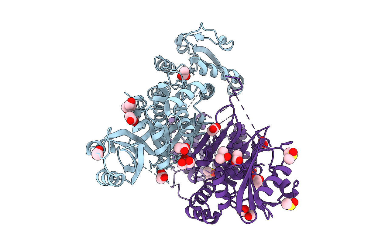
4PG5 image
Deposition Date 2014-05-01
Release Date 2015-05-06
Last Version Date 2023-09-27
Entry Detail
PDB ID:
4PG5
Keywords:
Title:
Crystal structure of S. aureus Homoserine Dehydrogenase at pH6.5
Biological Source:
Source Organism:
Host Organism:
Method Details:
Experimental Method:
Resolution:
2.20 Å
R-Value Free:
0.23
R-Value Work:
0.20
R-Value Observed:
0.20
Space Group:
P 21 21 21
Macromolecular Entities
Polymer Type:polypeptide(L)
Molecule:Homoserine dehydrogenase
Chain IDs:A, B
Chain Length:468
Number of Molecules:2
Biological Source:Staphylococcus aureus M1064
Primary Citation
Structural basis for the catalytic mechanism of homoserine dehydrogenase.
Acta Crystallogr.,Sect.D 71 1216 1225 (2015)
PMID: 25945586 DOI: 10.1107/S1399004715004617

Abstact

Homoserine dehydrogenase (HSD) is an oxidoreductase in the aspartic acid pathway. This enzyme coordinates a critical branch point of the metabolic pathway that leads to the synthesis of bacterial cell-wall components such as L-lysine and m-DAP in addition to other amino acids such as L-threonine, L-methionine and L-isoleucine. Here, a structural rationale for the hydride-transfer step in the reaction mechanism of HSD is reported. The structure of Staphylococcus aureus HSD was determined at different pH conditions to understand the basis for the enhanced enzymatic activity at basic pH. An analysis of the crystal structure revealed that Lys105, which is located at the interface of the catalytic and cofactor-binding sites, could mediate the hydride-transfer step of the reaction mechanism. The role of Lys105 was subsequently confirmed by mutational analysis. Put together, these studies reveal the role of conserved water molecules and a lysine residue in hydride transfer between the substrate and the cofactor.

Legend

Protein

Chemical

Disease

Primary Citation of related structures