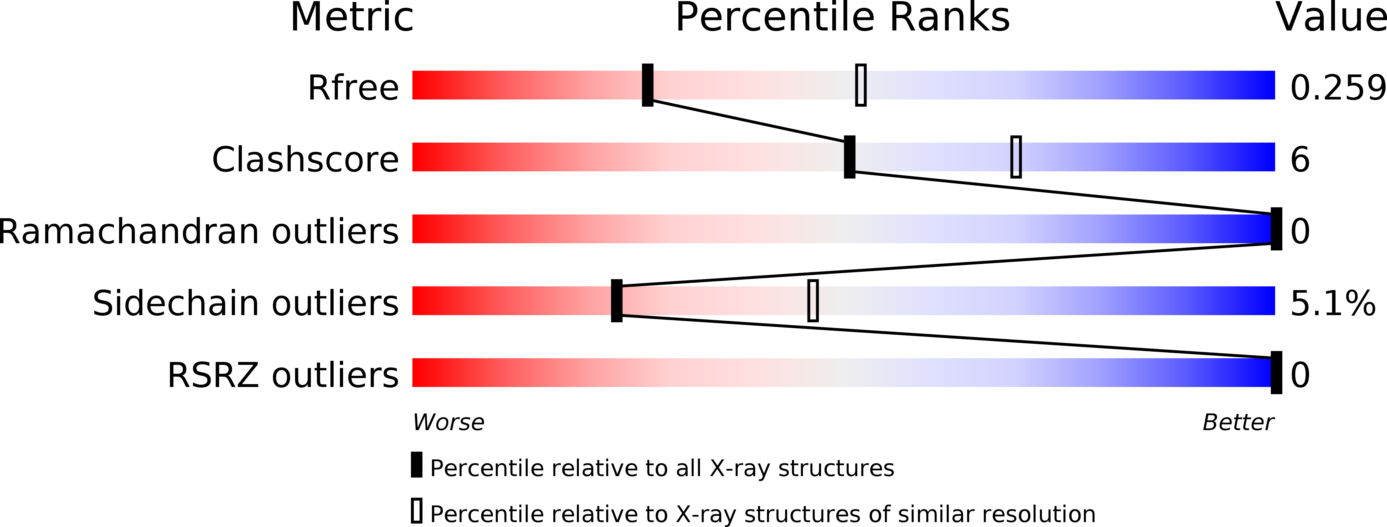
4PFE image
Deposition Date 2014-04-29
Release Date 2015-06-24
Last Version Date 2024-11-06
Entry Detail
PDB ID:
4PFE
Title:
Crystal structure of vsfGFP-0
Biological Source:
Source Organism:
Aequorea victoria (Taxon ID: 6100)
Vicugna pacos (Taxon ID: 30538)
Host Organism:
Method Details:
Experimental Method:
Resolution:
2.60 Å
R-Value Free:
0.25
R-Value Work:
0.19
R-Value Observed:
0.20
Space Group:
P 41 21 2
Macromolecular Entities
Structures with similar UniProt ID
Protein Blast
Polymer Type:polypeptide(L)
Molecule:Green fluorescent protein
Gene (Uniprot):GFP
Chain IDs:A, B
Chain Length:345
Number of Molecules:2
Biological Source:Aequorea victoria, Vicugna pacos
Modified Residue
Compound ID Chain ID Parent Comp ID Details 2D Image
CRO A SER chromophore
Primary Citation
Rational Structure-Based Design of Bright GFP-Based Complexes with Tunable Dimerization.
Angew.Chem.Int.Ed.Engl. 54 13952 13956 (2015)
PMID: 26447926 DOI: 10.1002/anie.201506686

Abstact

Fluorescent proteins are transformative tools; thus, any brightness increase is a welcome improvement. We invented the "vGFP strategy" based on structural analysis of GFP bound to a single-domain antibody, predicting tunable dimerization, enhanced brightness (ca. 50%), and improved pH resistance. We verified all of these predictions using biochemistry, crystallography, and single-molecule studies. We applied the vsfGFP proteins in three diverse scenarios: single-step immunofluorescence in vitro (3× brighter due to dimerization); expression in bacteria and human cells in vivo (1.5× brighter); and protein fusions showing better pH resistance in human cells in vivo. The vGFP strategy thus allows upgrading of existing applications, is applicable to other fluorescent proteins, and suggests a method for tuning dimerization of arbitrary proteins and optimizing protein properties in general.

Legend

Protein

Chemical

Disease

Primary Citation of related structures