4PD3 image
Deposition Date 2014-04-17
Release Date 2014-11-12
Last Version Date 2023-12-27
Entry Detail
PDB ID:
4PD3
Title:
Crystal Structure of Rigor-Like Human Nonmuscle Myosin-2B
Biological Source:
Source Organism:
Host Organism:
Method Details:
Experimental Method:
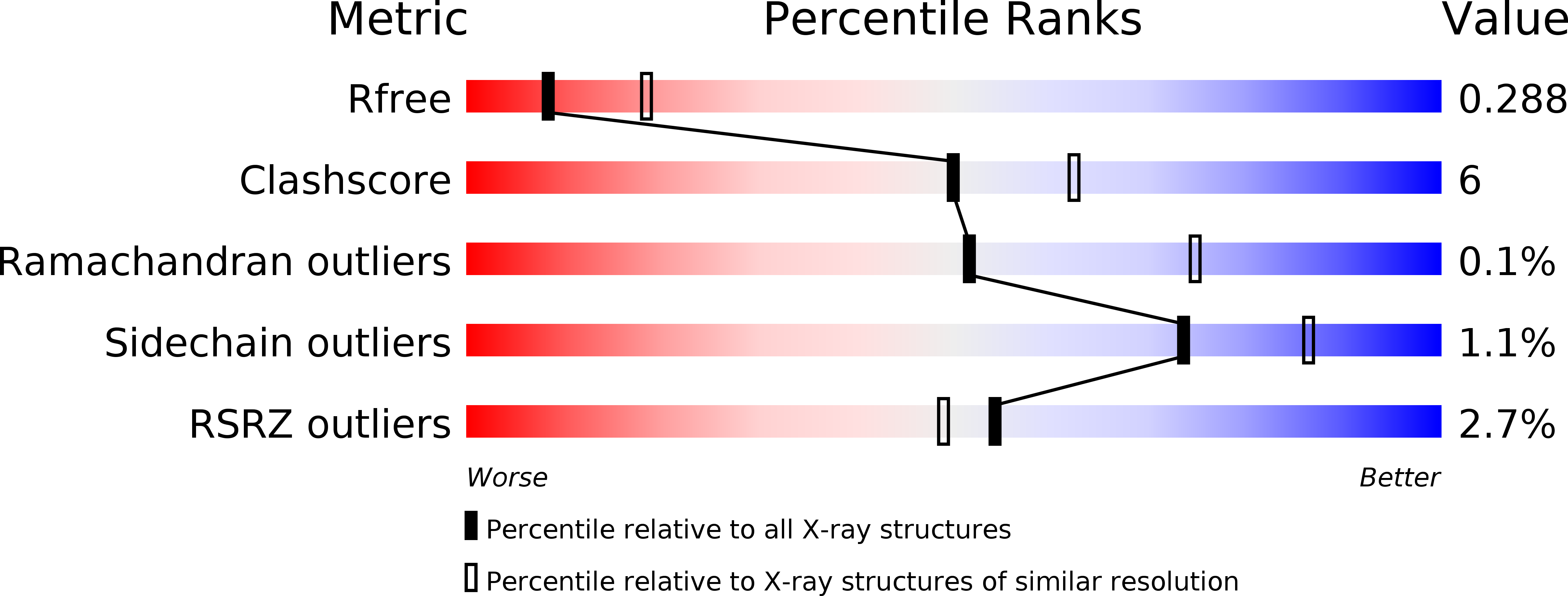
Resolution:
2.84 Å
R-Value Free:
0.28
R-Value Work:
0.25
R-Value Observed:
0.25
Space Group:
P 1 21 1
Macromolecular Entities
Protein Blast
Polymer Type:polypeptide(L)
Molecule:Nonmuscle myosin heavy chain B, Alpha-actinin A Chimera Protein
Gene (Uniprot):abpA, MYH10
Chain IDs:A, B
Chain Length:1032
Number of Molecules:2
Biological Source:Homo sapiens, Dictyostelium discoideum
Primary Citation
Crystal structure of the rigor-like human non-muscle myosin-2 motor domain.
Febs Lett. 588 4754 4760 (2014)
PMID: 25451231 DOI: 10.1016/j.febslet.2014.11.007

Abstact

We determined the crystal structure of the motor domain of human non-muscle myosin 2B (NM-2B) in a nucleotide-free state and at a resolution of 2.8 Å. The structure shows the motor domain with an open active site and the large cleft that divides the 50 kDa domain in a closed state. Compared to other rigor-like myosin motor domain structures, our structure shows subtle but significant conformational changes in regions important for actin binding and mechanochemical coupling. Moreover, our crystal structure helps to rationalize the impact of myosin, heavy chain 9 (MYH9)-related disease mutations Arg709Cys and Arg709His on the kinetic and functional properties of NM-2B and of the closely related non-muscle myosin 2A (NM-2A).

Legend

Protein

Chemical

Disease

Primary Citation of related structures