4P9A image
Entry Detail
PDB ID:
4P9A
Keywords:
Title:
X-ray Crystal Structure of PA protein from Influenza strain H7N9
Biological Source:
Source Organism:
Host Organism:
PDB Version:
Deposition Date:
2014-04-02
Release Date:
2014-07-30
Method Details:
Experimental Method:
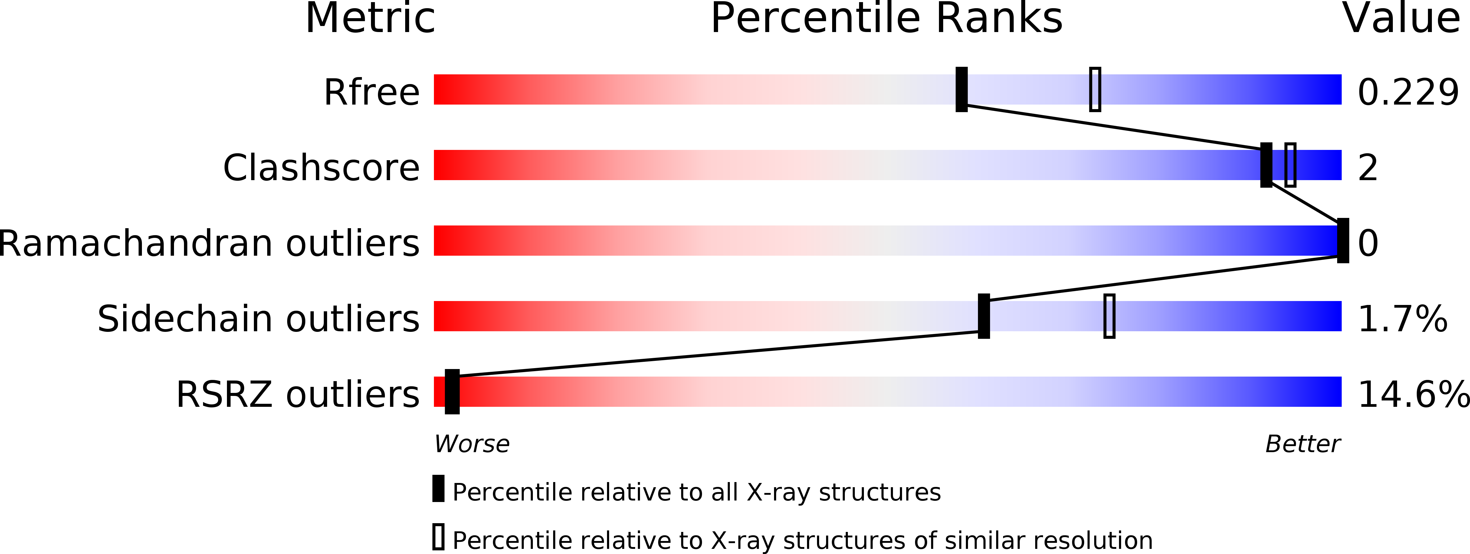
Resolution:
2.20 Å
R-Value Free:
0.22
R-Value Work:
0.20
R-Value Observed:
0.20
Space Group:
P 65 2 2
Macromolecular Entities
Polymer Type:polypeptide(L)
Description:Polymerase PA
Chain IDs:A
Chain Length:464
Number of Molecules:1
Biological Source:Influenza A virus
Ligand Molecules
Primary Citation
Structural analysis of H1N1 and H7N9 influenza A virus PA in the absence of PB1.
Sci Rep 4 5944 5944 (2014)
PMID: 25089892 DOI: 10.1038/srep05944

Abstact

Influenza A viruses cause the respiratory illness influenza, which can be mild to fatal depending on the strain and host immune response. The flu polymerase acidic (PA), polymerase basic 1 (PB1), and polymerase basic 2 (PB2) proteins comprise the RNA-dependent RNA polymerase complex responsible for viral genome replication. The first crystal structures of the C-terminal domain of PA (PA-CTD) in the absence of PB1-derived peptides show a number of structural changes relative to the previously reported PB1-peptide bound structures. The human A/WSN/1933 (H1N1) and avian A/Anhui1/2013 (H7N9) strain PA-CTD proteins exhibit the same global topology as other strains in the absence of PB1, but differ extensively in the PB1 binding pocket including a widening of the binding groove and the unfolding of a β-turn. Both PA-CTD proteins exhibited a significant increase in thermal stability in the presence of either a PB1-derived peptide or a previously reported inhibitor in differential scanning fluorimetry assays. These structural changes demonstrate plasticity in the PA-PB1 binding interface which may be exploited in the development of novel therapeutics.

Legend

Protein

Chemical

Disease

Primary Citation of related structures