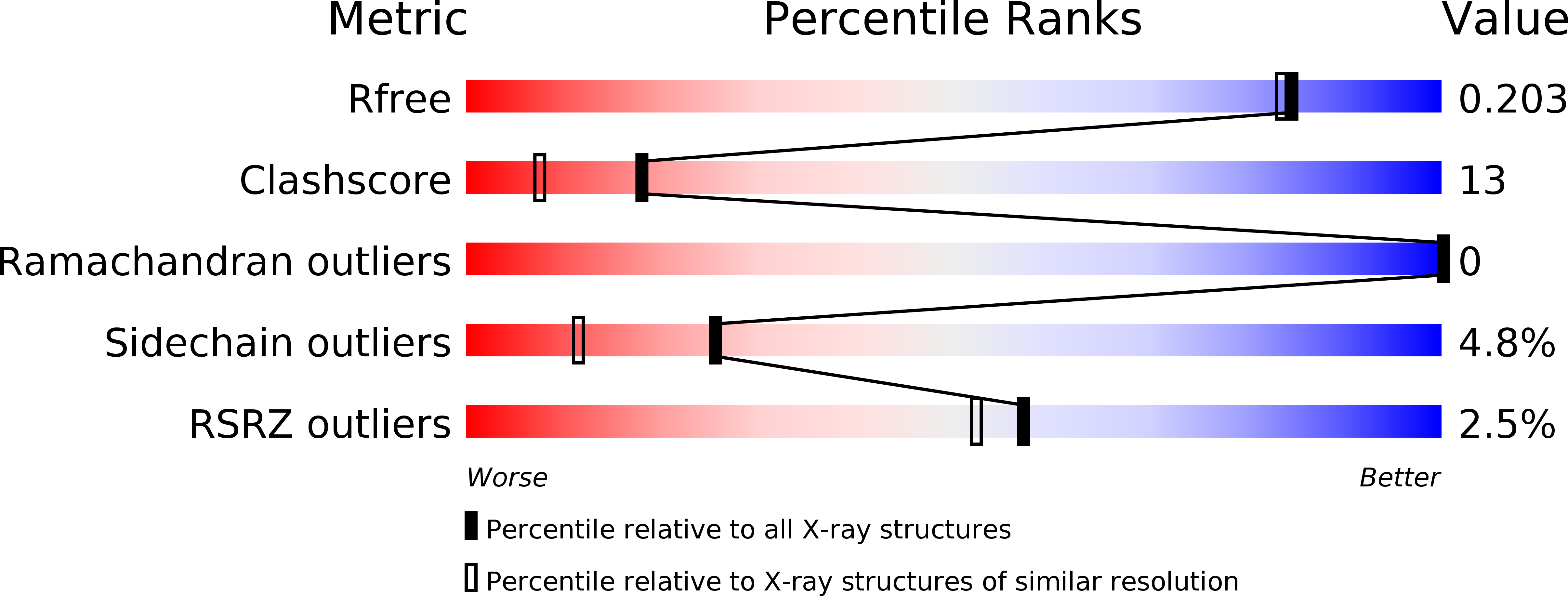
4P8S image
Deposition Date 2014-04-01
Release Date 2014-04-09
Last Version Date 2024-10-16
Entry Detail
PDB ID:
4P8S
Title:
Crystal structure of Nogo-receptor-2
Biological Source:
Source Organism:
Host Organism:
Method Details:
Experimental Method:
Resolution:
1.80 Å
R-Value Free:
0.20
R-Value Work:
0.17
R-Value Observed:
0.17
Space Group:
P 21 21 21
Macromolecular Entities
Polymer Type:polypeptide(L)
Molecule:Reticulon-4 receptor-like 2
Gene (Uniprot):Rtn4rl2
Chain IDs:A
Chain Length:282
Number of Molecules:1
Biological Source:Rattus norvegicus
Ligand Molecules
Primary Citation
Crystal structure of the Nogo-receptor-2.
Protein Sci. . 684 689 (2011)
PMID: 21308849 DOI: 10.1002/pro.597

Abstact

The inhibition of axon regeneration upon mechanical injury is dependent on interactions between Nogo receptors (NgRs) and their myelin-derived ligands. NgRs are composed of a leucine-rich repeat (LRR) region, thought to be structurally similar among the different isoforms of the receptor, and a divergent "stalk" region. It has been shown by others that the LRR and stalk regions of NgR1 and NgR2 have distinct roles in conferring binding affinity to the myelin associated glycoprotein (MAG) in vivo. Here, we show that purified recombinant full length NgR1 and NgR2 maintain significantly higher binding affinity for purified MAG as compared to the isolated LRR region of either NgR1 or NgR2. We also present the crystal structure of the LRR and part of the stalk regions of NgR2 and compare it to the previously reported NgR1 structure with respect to the distinct signaling properties of the two receptor isoforms.

Legend

Protein

Chemical

Disease

Primary Citation of related structures