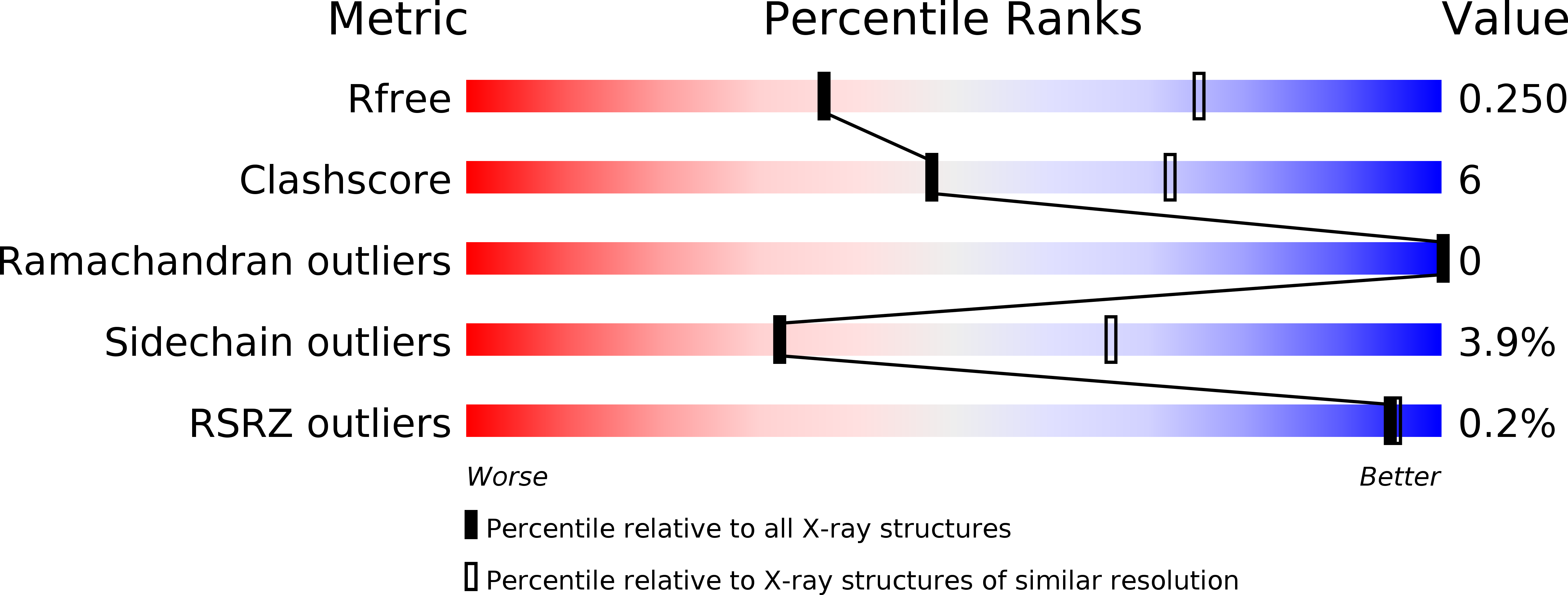
4P76 image
Deposition Date 2014-03-26
Release Date 2015-04-29
Last Version Date 2024-10-23
Entry Detail
PDB ID:
4P76
Title:
Cellular response to a crystal-forming protein
Biological Source:
Source Organism:
Favia favus (Taxon ID: 102203)
Host Organism:
Method Details:
Experimental Method:
Resolution:
2.90 Å
R-Value Free:
0.24
R-Value Work:
0.17
R-Value Observed:
0.17
Space Group:
P 21 21 2
Macromolecular Entities
Polymer Type:polypeptide(L)
Molecule:Photoconvertible fluorescent protein
Gene (Uniprot):KikGR
Mutations:Q198S
Chain IDs:A, B
Chain Length:228
Number of Molecules:2
Biological Source:Favia favus
Modified Residue
Compound ID Chain ID Parent Comp ID Details 2D Image
CR8 A HIS chromophore
Ligand Molecules
Primary Citation
A diffraction-quality protein crystal processed as an autophagic cargo
Mol.Cell 58 186 193 (2015)
PMID: 25773597 DOI: 10.1016/j.molcel.2015.02.007

Abstact

Crystallization of proteins may occur in the cytosol of a living cell, but how a cell responds to intracellular protein crystallization remains unknown. We developed a variant of coral fluorescent protein that forms diffraction-quality crystals within mammalian cells. This expression system allowed the direct determination of its crystal structure at 2.9 Å, as well as observation of the crystallization process and cellular responses. The micron-sized crystal, which emerged rapidly, was a pure assembly of properly folded β-barrels and was recognized as an autophagic cargo that was transferred to lysosomes via a process involving p62 and LC3. Several lines of evidence indicated that autophagy was not required for crystal nucleation or growth. These findings demonstrate that in vivo protein crystals can provide an experimental model to study chemical catalysis. This knowledge may be beneficial for structural biology studies on normal and disease-related protein aggregation.

Legend

Protein

Chemical

Disease

Primary Citation of related structures