Deposition Date
2014-03-20
Release Date
2014-08-13
Last Version Date
2023-12-20
Entry Detail
PDB ID:
4P5Z
Keywords:
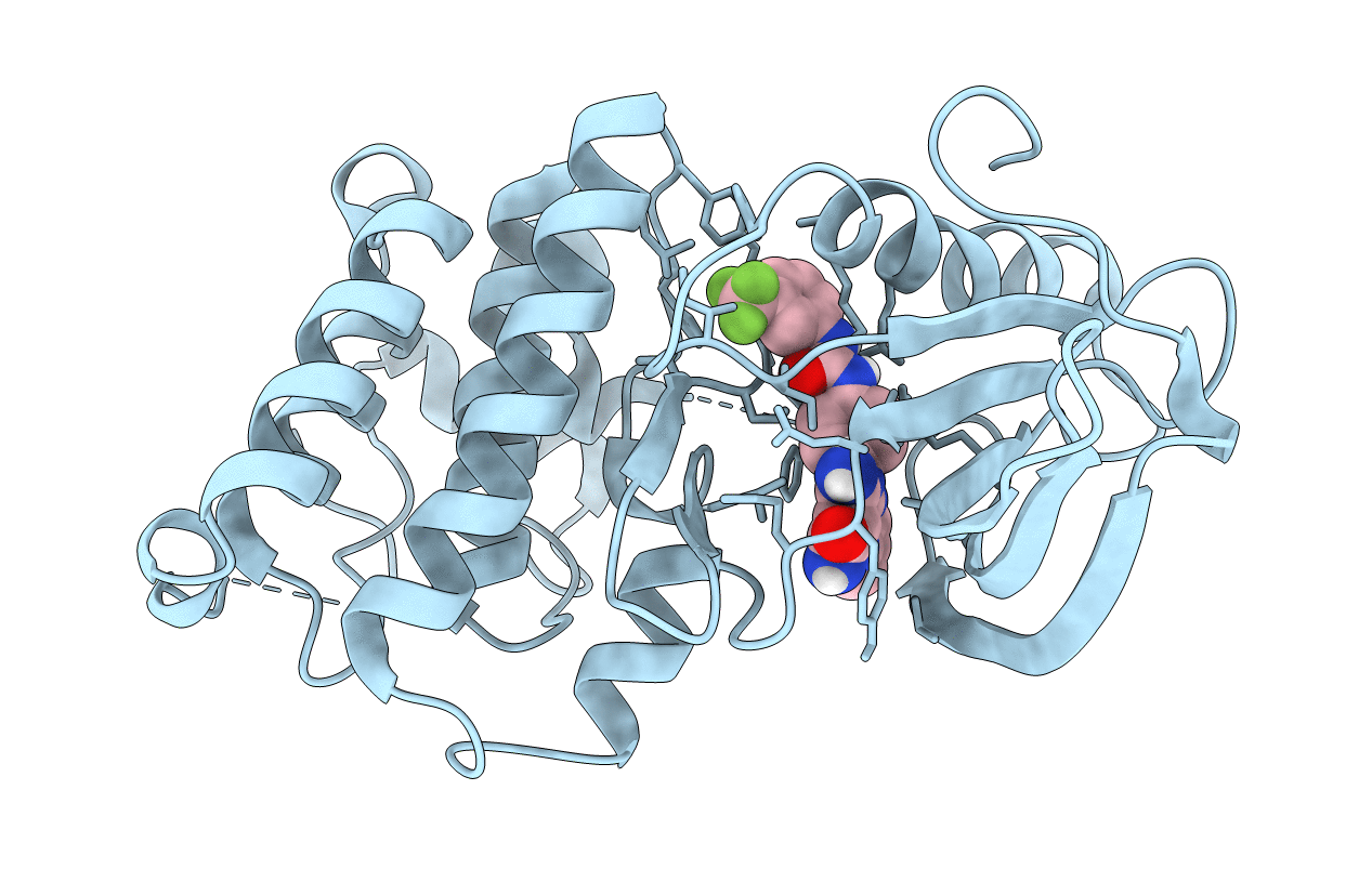
Title:
Human EphA3 Kinase domain in complex with quinoxaline derivatives
Biological Source:
Source Organism(s):
Homo sapiens (Taxon ID: 9606)
Expression System(s):
Method Details:
Experimental Method:
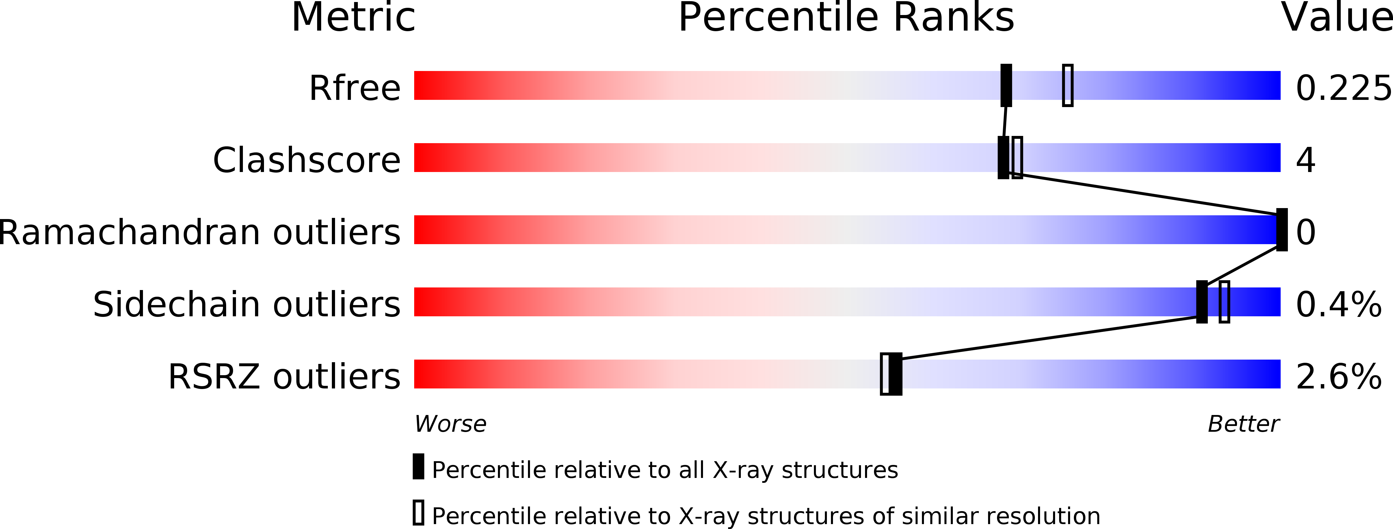
Resolution:
2.00 Å
R-Value Free:
0.21
R-Value Work:
0.17
R-Value Observed:
0.18
Space Group:
P 1 21 1