Deposition Date
2014-03-20
Release Date
2014-11-26
Last Version Date
2023-12-20
Entry Detail
PDB ID:
4P5X
Keywords:
Title:
Structure of the N-terminal domain of the human mitochondrial aspartate/glutamate carrier Aralar in the calcium-bound state
Biological Source:
Source Organism(s):
Homo sapiens (Taxon ID: 9606)
Expression System(s):
Method Details:
Experimental Method:
Resolution:
2.26 Å
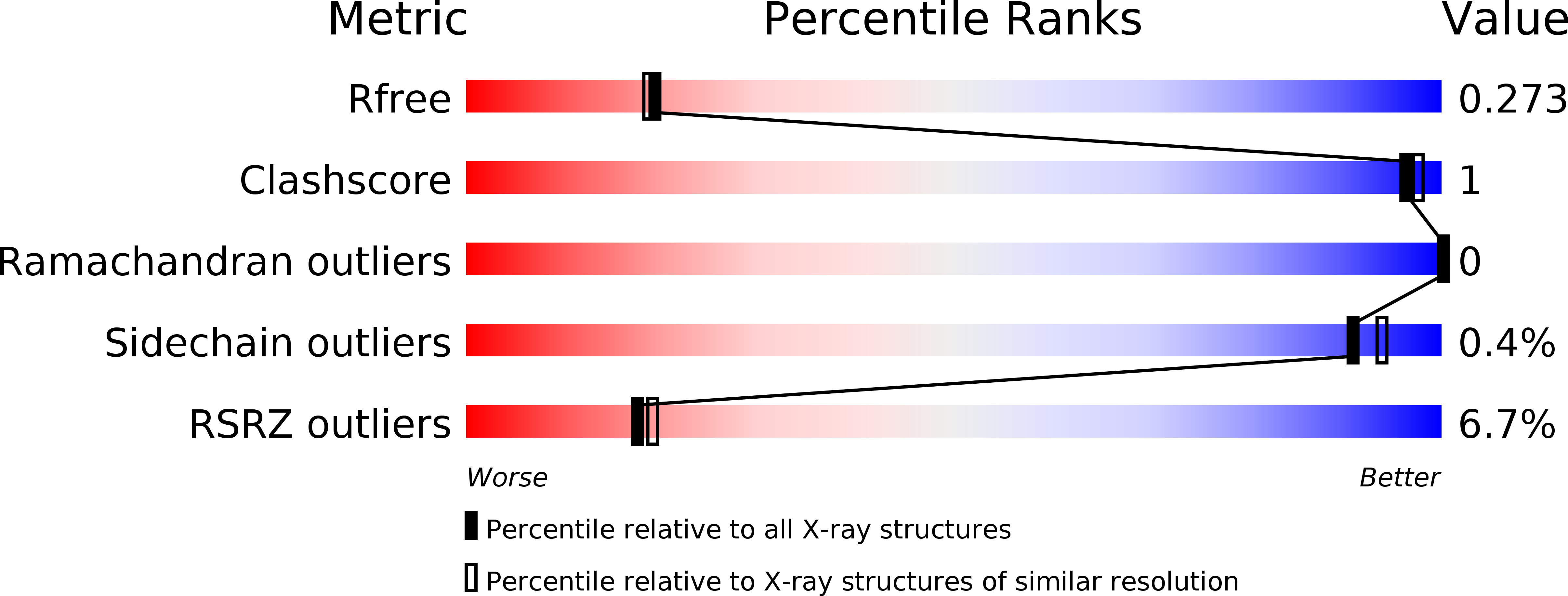
R-Value Free:
0.27
R-Value Work:
0.23
R-Value Observed:
0.23
Space Group:
P 31 2 1