Deposition Date
2014-03-10
Release Date
2014-06-18
Last Version Date
2023-10-25
Entry Detail
PDB ID:
4P3Y
Keywords:
Title:
Crystal structure of Acinetobacter baumannii DsbA in complex with EF-Tu
Biological Source:
Source Organism(s):
Acinetobacter baumannii AYE (Taxon ID: 509173)
Escherichia coli BL21(DE3) (Taxon ID: 469008)
Escherichia coli BL21(DE3) (Taxon ID: 469008)
Expression System(s):
Method Details:
Experimental Method:
Resolution:
2.15 Å
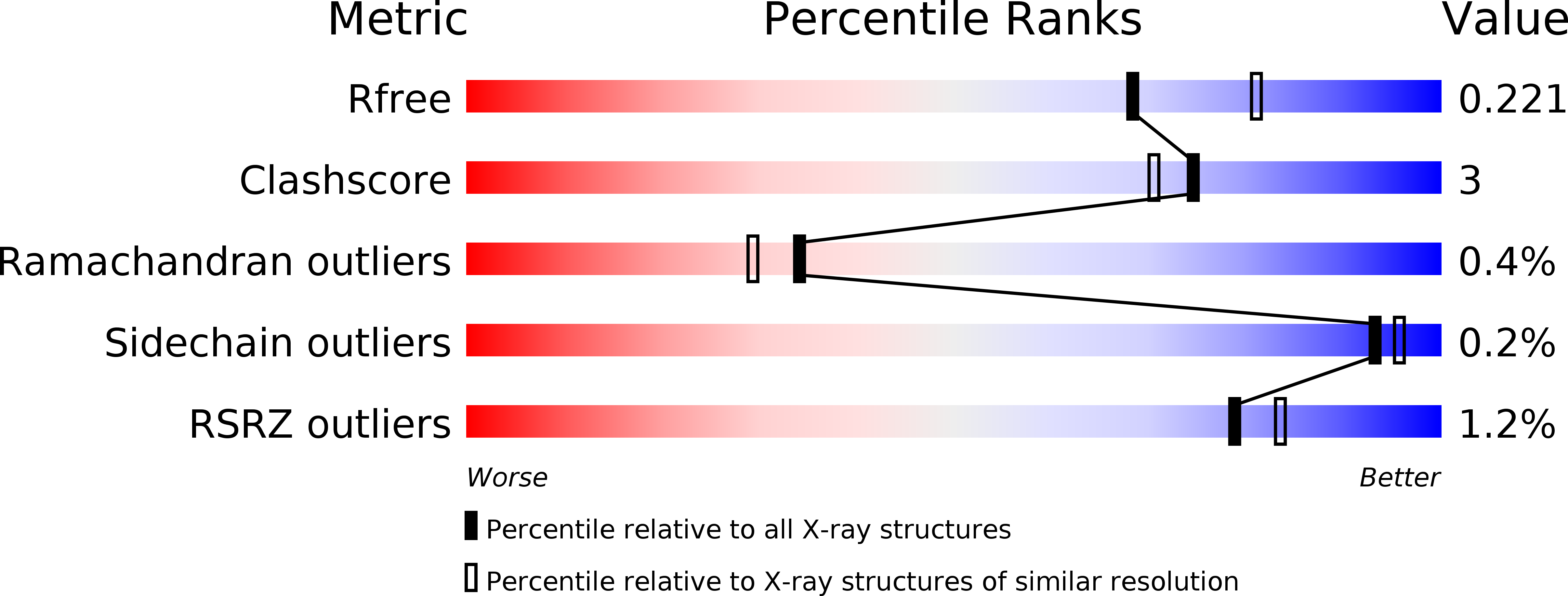
R-Value Free:
0.21
R-Value Work:
0.16
R-Value Observed:
0.17
Space Group:
P 21 21 21