4P29 image
Deposition Date 2014-03-02
Release Date 2015-03-04
Last Version Date 2024-10-16
Entry Detail
PDB ID:
4P29
Keywords:
Title:
Crystal structure of the LpoA N-terminal domain from Haemophilus influenzae
Biological Source:
Source Organism:
Host Organism:
Method Details:
Experimental Method:
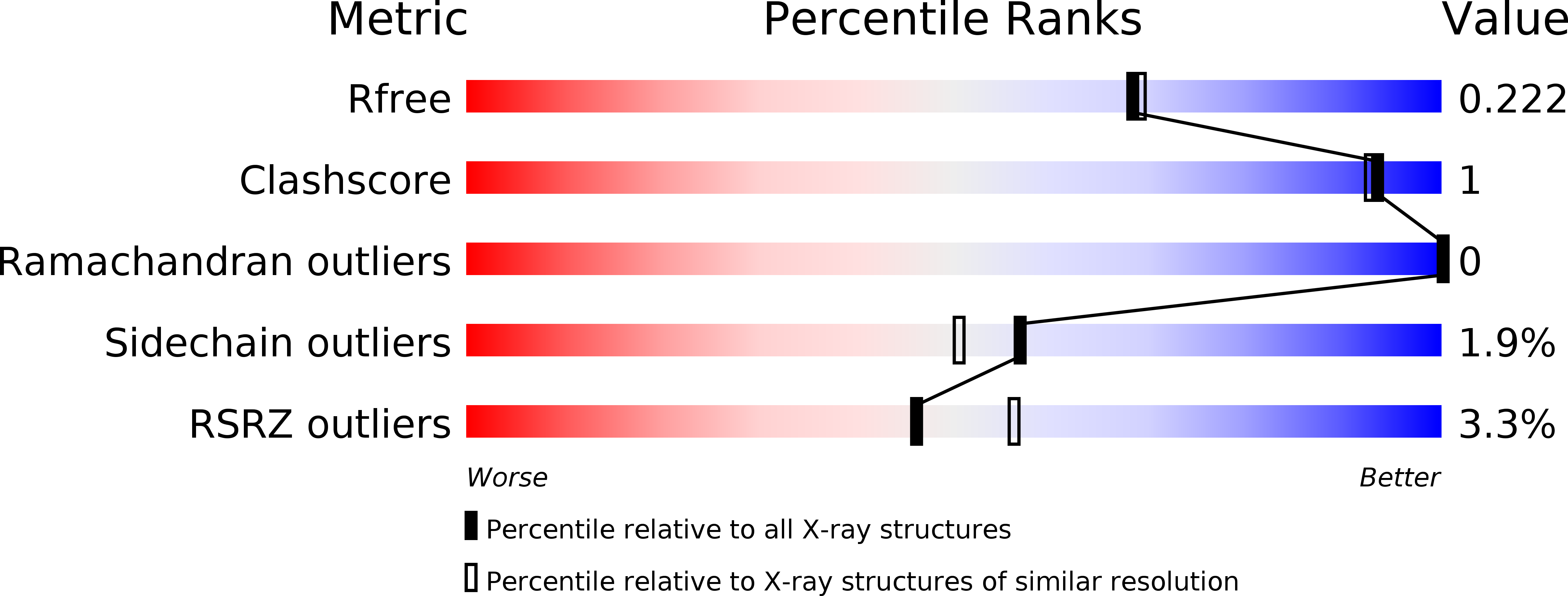
Resolution:
1.95 Å
R-Value Free:
0.21
R-Value Work:
0.18
R-Value Observed:
0.18
Space Group:
P 21 21 21
Macromolecular Entities
Structures with similar UniProt ID
Protein Blast
Polymer Type:polypeptide(L)
Molecule:LpoA
Gene (Uniprot):lpoA
Chain IDs:A, B
Chain Length:231
Number of Molecules:2
Biological Source:Haemophilus influenzae
Modified Residue
Compound ID Chain ID Parent Comp ID Details 2D Image
MSE A MET modified residue
Primary Citation
Structural analyses of theHaemophilus influenzaepeptidoglycan synthase activator LpoA suggest multiple conformations in solution.
J.Biol.Chem. 292 17626 17642 (2017)
PMID: 28887305 DOI: 10.1074/jbc.M117.804997

Abstact

In many Gram-negative bacteria, the peptidoglycan synthase PBP1A requires the outer membrane lipoprotein LpoA for constructing a functional peptidoglycan required for bacterial viability. Previously, we have shown that the C-terminal domain of Haemophilus influenzae LpoA (HiLpoA) has a highly conserved, putative substrate-binding cleft between two α/β lobes. Here, we report a 2.0 Å resolution crystal structure of the HiLpoA N-terminal domain. Two subdomains contain tetratricopeptide-like motifs that form a concave groove, but their relative orientation differs by ∼45° from that observed in an NMR structure of the Escherichia coli LpoA N domain. We also determined three 2.0-2.8 Å resolution crystal structures containing four independent full-length HiLpoA molecules. In contrast to an elongated model previously suggested for E. coli LpoA, each HiLpoA formed a U-shaped structure with a different C-domain orientation. This resulted from both N-domain twisting and rotation of the C domain (up to 30°) at the end of the relatively immobile interdomain linker. Moreover, a previously predicted hinge between the lobes of the LpoA C domain exhibited variations of up to 12°. Small-angle X-ray scattering data revealed excellent agreement with a model calculated by normal mode analysis from one of the full-length HiLpoA molecules but even better agreement with an ensemble of this molecule and two of the partially extended normal mode analysis-predicted models. The different LpoA structures helped explain how an outer membrane-anchored LpoA can either withdraw from or extend toward the inner membrane-bound PBP1A through peptidoglycan gaps and hence regulate the synthesis of peptidoglycan necessary for bacterial viability.

Legend

Protein

Chemical

Disease

Primary Citation of related structures
Feedback Form
Name
Email
Institute
Feedback