4P1Y image
Deposition Date 2014-02-28
Release Date 2014-10-01
Last Version Date 2023-12-27
Entry Detail
PDB ID:
4P1Y
Keywords:
Title:
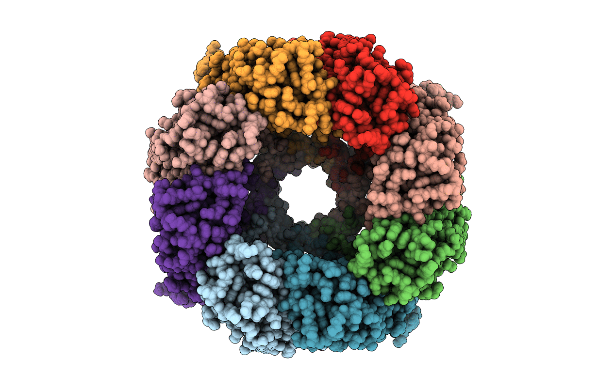
Crystal structure of staphylococcal gamma-hemolysin prepore
Biological Source:
Source Organism:
Host Organism:
Method Details:
Experimental Method:
Resolution:
2.99 Å
R-Value Free:
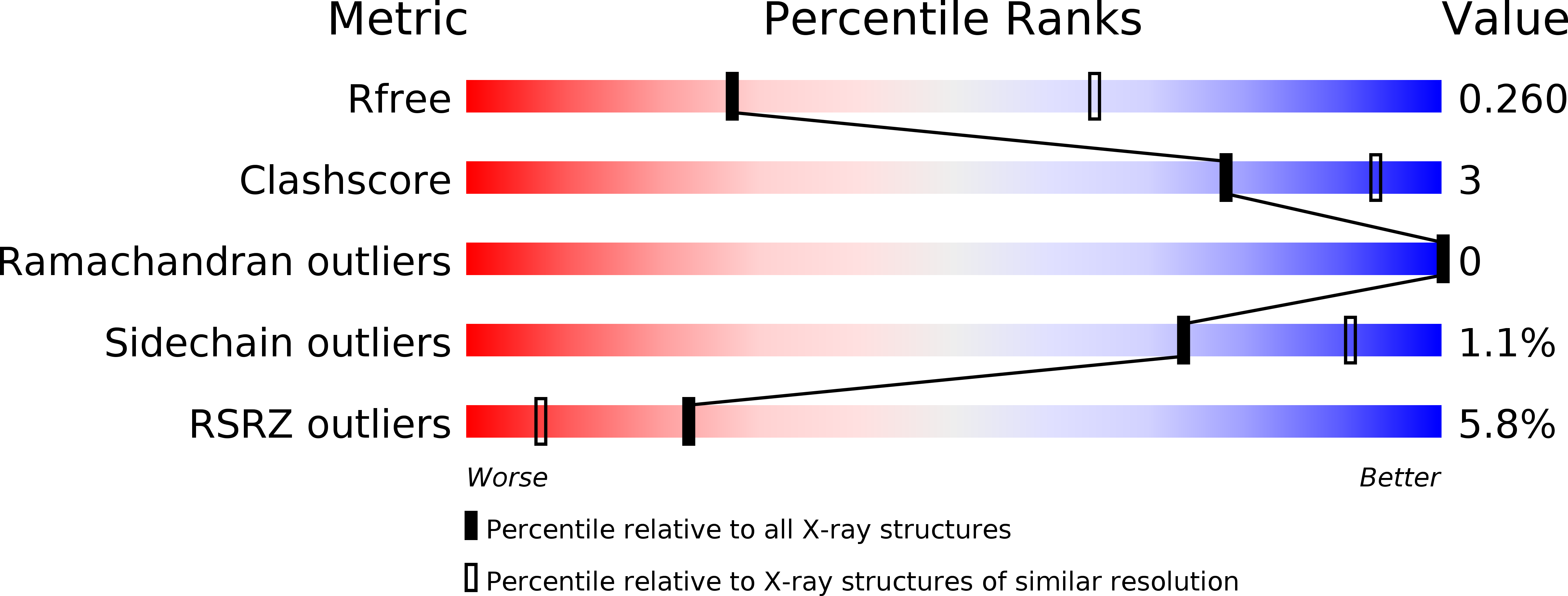
0.25
R-Value Work:
0.23
R-Value Observed:
0.23
Space Group:
C 1 2 1
Macromolecular Entities
Polymer Type:polypeptide(L)
Molecule:Gamma-hemolysin component B
Mutations:W177A R198A
Chain IDs:A, C, E, G
Chain Length:309
Number of Molecules:4
Biological Source:Staphylococcus aureus
Polymer Type:polypeptide(L)
Molecule:Gamma-hemolysin component A
Gene (Uniprot):hlgA
Chain IDs:B, D, F, H
Chain Length:290
Number of Molecules:4
Biological Source:Staphylococcus aureus
Ligand Molecules
Primary Citation
Molecular basis of transmembrane beta-barrel formation of staphylococcal pore-forming toxins.
Nat Commun 5 4897 4897 (2014)
PMID: 25263813 DOI: 10.1038/ncomms5897

Abstact

Pathogenic bacteria secrete pore-forming toxins (PFTs) to attack target cells. PFTs are expressed as water-soluble monomeric proteins, which oligomerize into nonlytic prepore intermediates on the target cell membrane before forming membrane-spanning pores. Despite a wealth of biochemical data, the lack of high-resolution prepore structural information has hampered understanding of the β-barrel formation process. Here, we report crystal structures of staphylococcal γ-haemolysin and leucocidin prepores. The structures reveal a disordered bottom half of the β-barrel corresponding to the transmembrane region, and a rigid upper extramembrane half. Spectroscopic analysis of fluorescently labelled mutants confirmed that the prepore is distinct from the pore within the transmembrane region. Mutational analysis also indicates a pivotal role for the glycine residue located at the lipid-solvent interface as a 'joint' between the two halves of the β-barrel. These observations suggest a two-step transmembrane β-barrel pore formation mechanism in which the upper extramembrane and bottom transmembrane regions are formed independently.

Legend

Protein

Chemical

Disease

Primary Citation of related structures