4P1B image
Deposition Date 2014-02-25
Release Date 2014-10-01
Last Version Date 2023-09-27
Entry Detail
PDB ID:
4P1B
Keywords:
Title:
CRYSTAL STRUCTURE OF THE TOLUENE 4-MONOOXYGENASE HYDROXYLASE-FERREDOXIN C7S E16C C84A C85A VARIANT ELECTRON-TRANSFER COMPLEX
Biological Source:
Source Organism:
Host Organism:
Method Details:
Experimental Method:
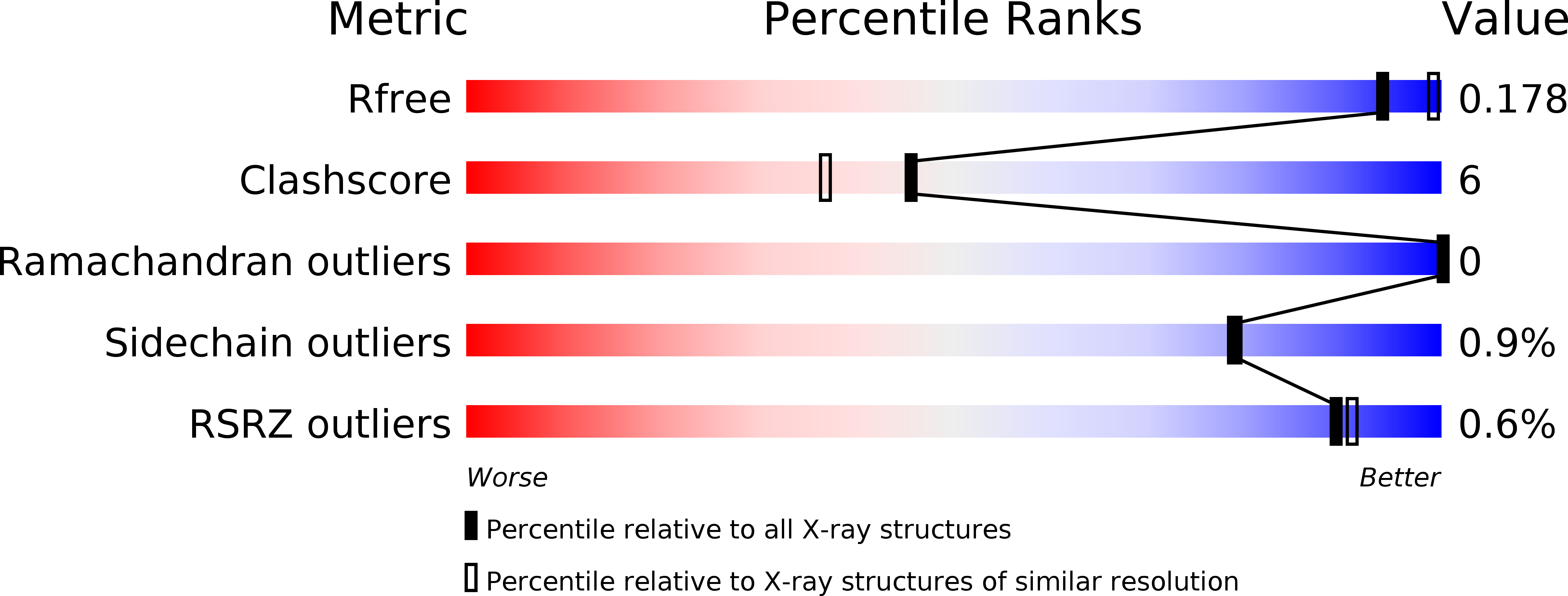
Resolution:
2.05 Å
R-Value Free:
0.17
R-Value Work:
0.14
R-Value Observed:
0.14
Space Group:
P 32 2 1
Macromolecular Entities
Structures with similar UniProt ID
Protein Blast
Polymer Type:polypeptide(L)
Molecule:Toluene-4-monooxygenase system protein A
Gene (Uniprot):tmoA
Mutagens:Stop 492
Chain IDs:A, D
Chain Length:490
Number of Molecules:2
Biological Source:Pseudomonas mendocina
Structures with similar UniProt ID
Protein Blast
Polymer Type:polypeptide(L)
Molecule:Toluene-4-monooxygenase system protein E
Gene (Uniprot):tmoE
Mutagens:Stop 307
Chain IDs:B, E
Chain Length:305
Number of Molecules:2
Biological Source:Pseudomonas mendocina
Structures with similar UniProt ID
Protein Blast
Polymer Type:polypeptide(L)
Molecule:Toluene-4-monooxygenase system protein B
Gene (Uniprot):tmoB
Chain IDs:C, F
Chain Length:82
Number of Molecules:2
Biological Source:Pseudomonas mendocina
Structures with similar UniProt ID
Protein Blast
Polymer Type:polypeptide(L)
Molecule:Toluene-4-monooxygenase system ferredoxin subunit
Gene (Uniprot):tmoC
Mutagens:C7S, E16C, C84A,C85A
Chain IDs:G (auth: H), H (auth: I)
Chain Length:111
Number of Molecules:2
Biological Source:Pseudomonas mendocina
Primary Citation
Structural basis for biomolecular recognition in overlapping binding sites in a diiron enzyme system.
Nat Commun 5 5009 5009 (2014)
PMID: 25248368 DOI: 10.1038/ncomms6009

Abstact

Productive biomolecular recognition requires exquisite control of affinity and specificity. Accordingly, nature has devised many strategies to achieve proper binding interactions. Bacterial multicomponent monooxygenases provide a fascinating example, where a diiron hydroxylase must reversibly interact with both ferredoxin and catalytic effector in order to achieve electron transfer and O2 activation during catalysis. Because these two accessory proteins have distinct structures, and because the hydroxylase-effector complex covers the entire surface closest to the hydroxylase diiron centre, how ferredoxin binds to the hydroxylase has been unclear. Here we present high-resolution structures of toluene 4-monooxygenase hydroxylase complexed with its electron transfer ferredoxin and compare them with the hydroxylase-effector structure. These structures reveal that ferredoxin or effector protein binding produce different arrangements of conserved residues and customized interfaces on the hydroxylase in order to achieve different aspects of catalysis.

Legend

Protein

Chemical

Disease

Primary Citation of related structures
Feedback Form
Name
Email
Institute
Feedback