4P0M image
Deposition Date 2014-02-21
Release Date 2014-03-12
Last Version Date 2023-12-27
Entry Detail
PDB ID:
4P0M
Keywords:
Title:
Crystal structure of an evolved putative penicillin-binding protein homolog, Rv2911, from Mycobacterium tuberculosis
Biological Source:
Source Organism:
Host Organism:
Method Details:
Experimental Method:
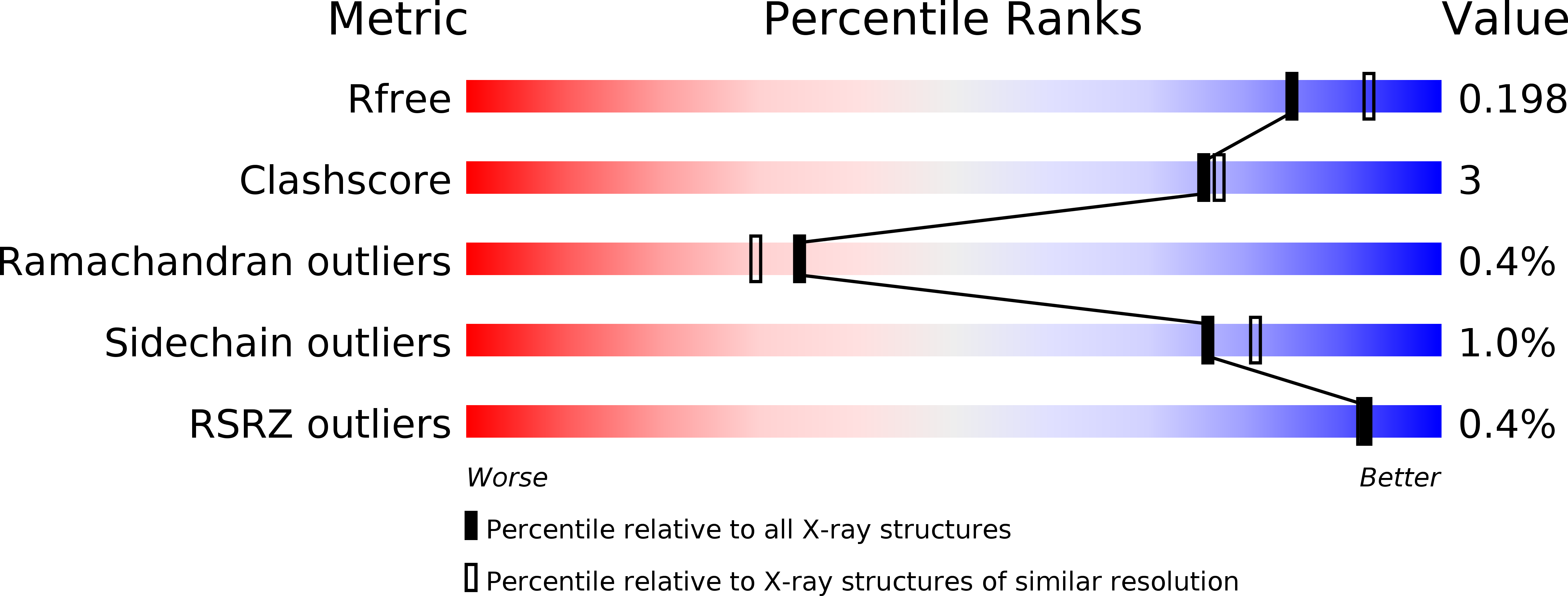
Resolution:
2.00 Å
R-Value Free:
0.19
R-Value Work:
0.17
R-Value Observed:
0.17
Space Group:
H 3 2
Macromolecular Entities
Polymer Type:polypeptide(L)
Molecule:D-alanyl-D-alanine carboxypeptidase
Gene (Uniprot):dacB
Mutations:A113V, T197I, N208D, G289D
Chain IDs:A
Chain Length:276
Number of Molecules:1
Biological Source:Mycobacterium tuberculosis
Modified Residue
Compound ID Chain ID Parent Comp ID Details 2D Image
MSE A MET modified residue
SEB A SER modified residue
Primary Citation
Subfamily-Specific Adaptations in the Structures of Two Penicillin-Binding Proteins from Mycobacterium tuberculosis.
Plos One 9 e116249 e116249 (2014)
PMID: 25551456 DOI: 10.1371/journal.pone.0116249

Abstact

Beta-lactam antibiotics target penicillin-binding proteins including several enzyme classes essential for bacterial cell-wall homeostasis. To better understand the functional and inhibitor-binding specificities of penicillin-binding proteins from the pathogen, Mycobacterium tuberculosis, we carried out structural and phylogenetic analysis of two predicted D,D-carboxypeptidases, Rv2911 and Rv3330. Optimization of Rv2911 for crystallization using directed evolution and the GFP folding reporter method yielded a soluble quadruple mutant. Structures of optimized Rv2911 bound to phenylmethylsulfonyl fluoride and Rv3330 bound to meropenem show that, in contrast to the nonspecific inhibitor, meropenem forms an extended interaction with the enzyme along a conserved surface. Phylogenetic analysis shows that Rv2911 and Rv3330 belong to different clades that emerged in Actinobacteria and are not represented in model organisms such as Escherichia coli and Bacillus subtilis. Clade-specific adaptations allow these enzymes to fulfill distinct physiological roles despite strict conservation of core catalytic residues. The characteristic differences include potential protein-protein interaction surfaces and specificity-determining residues surrounding the catalytic site. Overall, these structural insights lay the groundwork to develop improved beta-lactam therapeutics for tuberculosis.

Legend

Protein

Chemical

Disease

Primary Citation of related structures