4OVV image
Deposition Date 2014-01-14
Release Date 2014-09-03
Last Version Date 2023-12-27
Entry Detail
PDB ID:
4OVV
Title:
Crystal Structure of PI3Kalpha in complex with diC4-PIP2
Biological Source:
Source Organism:
Homo sapiens (Taxon ID: 9606)
Host Organism:
Method Details:
Experimental Method:
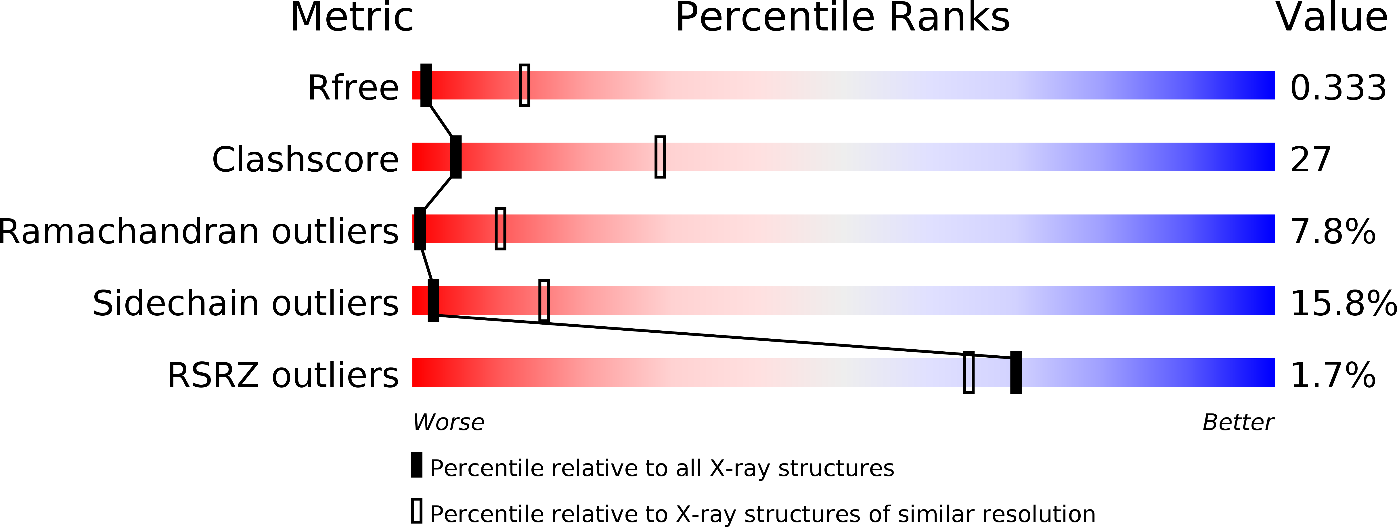
Resolution:
3.50 Å
R-Value Free:
0.33
R-Value Work:
0.23
R-Value Observed:
0.24
Space Group:
P 21 21 21
Macromolecular Entities
Polymer Type:polypeptide(L)
Molecule:Phosphatidylinositol 4,5-bisphosphate 3-kinase catalytic subunit alpha isoform
Gene (Uniprot):PIK3CA
Chain IDs:A
Chain Length:1096
Number of Molecules:1
Biological Source:Homo sapiens
Polymer Type:polypeptide(L)
Molecule:Phosphatidylinositol 3-kinase regulatory subunit alpha
Gene (Uniprot):PIK3R1
Chain IDs:B
Chain Length:279
Number of Molecules:1
Biological Source:Homo sapiens
Primary Citation
Structural basis of nSH2 regulation and lipid binding in PI3K alpha.
Oncotarget 5 5198 5208 (2014)
PMID: 25105564

Abstact

We report two crystal structures of the wild-type phosphatidylinositol 3-kinase α (PI3Kα) heterodimer refined to 2.9 Å and 3.4 Å resolution: the first as the free enzyme, the second in complex with the lipid substrate, diC4-PIP₂, respectively. The first structure shows key interactions of the N-terminal SH2 domain (nSH2) and iSH2 with the activation loop that suggest a mechanism by which the enzyme is inhibited in its basal state. In the second structure, the lipid substrate binds in a positively charged pocket adjacent to the ATP-binding site, bordered by the P-loop, the activation loop and the iSH2 domain. An additional lipid-binding site was identified at the interface of the ABD, iSH2 and kinase domains. The ability of PI3Kα to bind an additional PIP₂ molecule was confirmed in vitro by fluorescence quenching experiments. The crystal structures reveal key differences in the way the nSH2 domain interacts with wild-type p110α and with the oncogenic mutant p110αH1047R. Increased buried surface area and two unique salt-bridges observed only in the wild-type structure suggest tighter inhibition in the wild-type PI3Kα than in the oncogenic mutant. These differences may be partially responsible for the increased basal lipid kinase activity and increased membrane binding of the oncogenic mutant.

Legend

Protein

Chemical

Disease

Primary Citation of related structures