4OQW image
Entry Detail
PDB ID:
4OQW
Title:
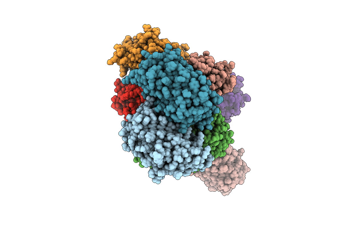
Crystal structure of mCardinal far-red fluorescent protein
Biological Source:
Source Organism:
Host Organism:
PDB Version:
Deposition Date:
2014-02-10
Release Date:
2014-03-12
Method Details:
Experimental Method:
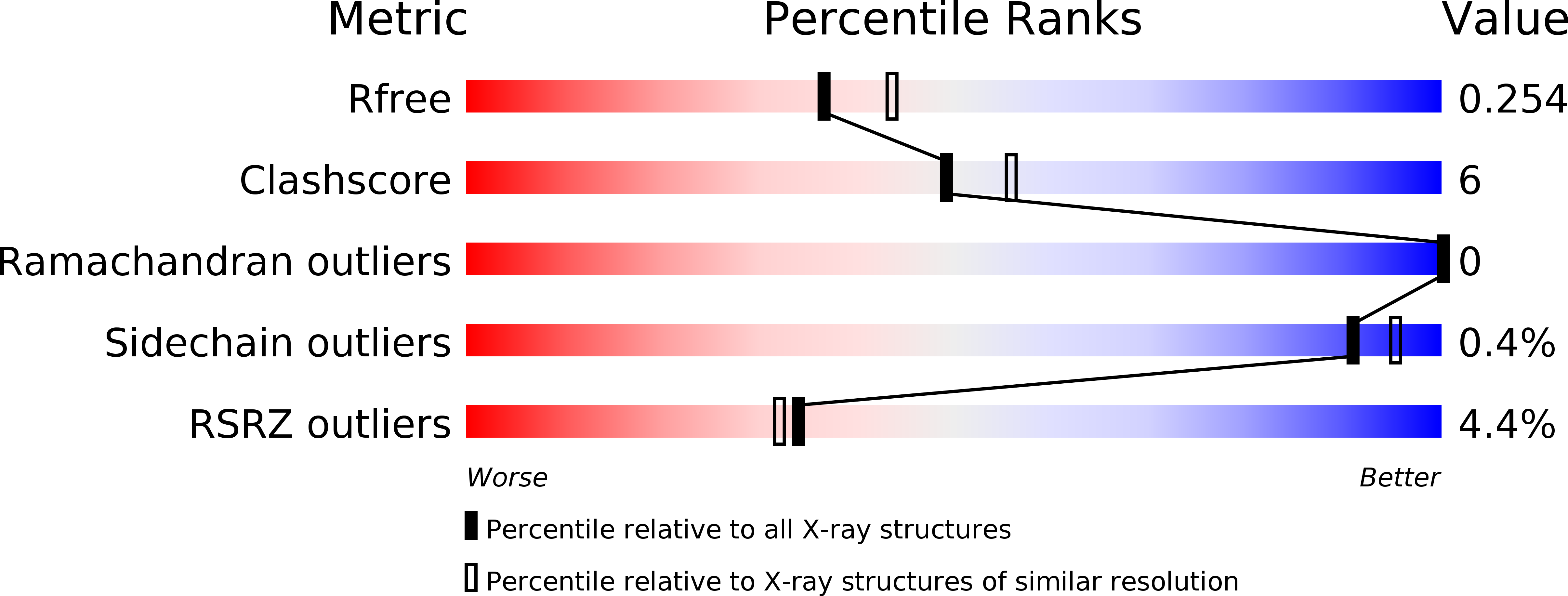
Resolution:
2.21 Å
R-Value Free:
0.25
R-Value Work:
0.22
R-Value Observed:
0.22
Space Group:
P 1 21 1
Macromolecular Entities
Polymer Type:polypeptide(L)
Description:Fluorescent protein FP480
Chain IDs:A, B, C, D, E, F, G, H
Chain Length:230
Number of Molecules:8
Biological Source:Entacmaea quadricolor
Modified Residue
Compound ID Chain ID Parent Comp ID Details 2D Image
MLY A LYS N-DIMETHYL-LYSINE
NRQ A MET ?
Primary Citation
Non-invasive intravital imaging of cellular differentiation with a bright red-excitable fluorescent protein.
Nat.Methods 11 572 578 (2014)
PMID: 24633408 DOI: 10.1038/nmeth.2888

Abstact

A method for non-invasive visualization of genetically labeled cells in animal disease models with micrometer-level resolution would greatly facilitate development of cell-based therapies. Imaging of fluorescent proteins (FPs) using red excitation light in the 'optical window' above 600 nm is one potential method for visualizing implanted cells. However, previous efforts to engineer FPs with peak excitation beyond 600 nm have resulted in undesirable reductions in brightness. Here we report three new red-excitable monomeric FPs obtained by structure-guided mutagenesis of mNeptune. Two of these, mNeptune2 and mNeptune2.5, demonstrate improved maturation and brighter fluorescence than mNeptune, whereas the third, mCardinal, has a red-shifted excitation spectrum without reduction in brightness. We show that mCardinal can be used to non-invasively and longitudinally visualize the differentiation of myoblasts into myocytes in living mice with high anatomical detail.

Legend

Protein

Chemical

Disease

Primary Citation of related structures