4OQO image
Deposition Date 2014-02-10
Release Date 2014-03-12
Last Version Date 2024-10-30
Entry Detail
PDB ID:
4OQO
Keywords:
Title:
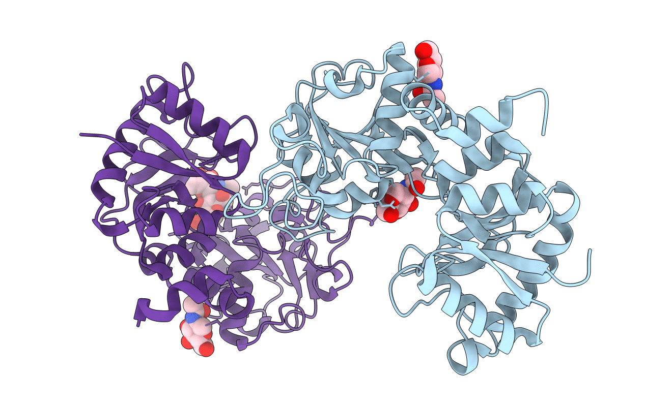
Crystal structure of the tryptic generated iron-free C-lobe of bovine Lactoferrin at 2.42 Angstrom resolution
Biological Source:
Source Organism:
Bos taurus (Taxon ID: 9913)
Method Details:
Experimental Method:
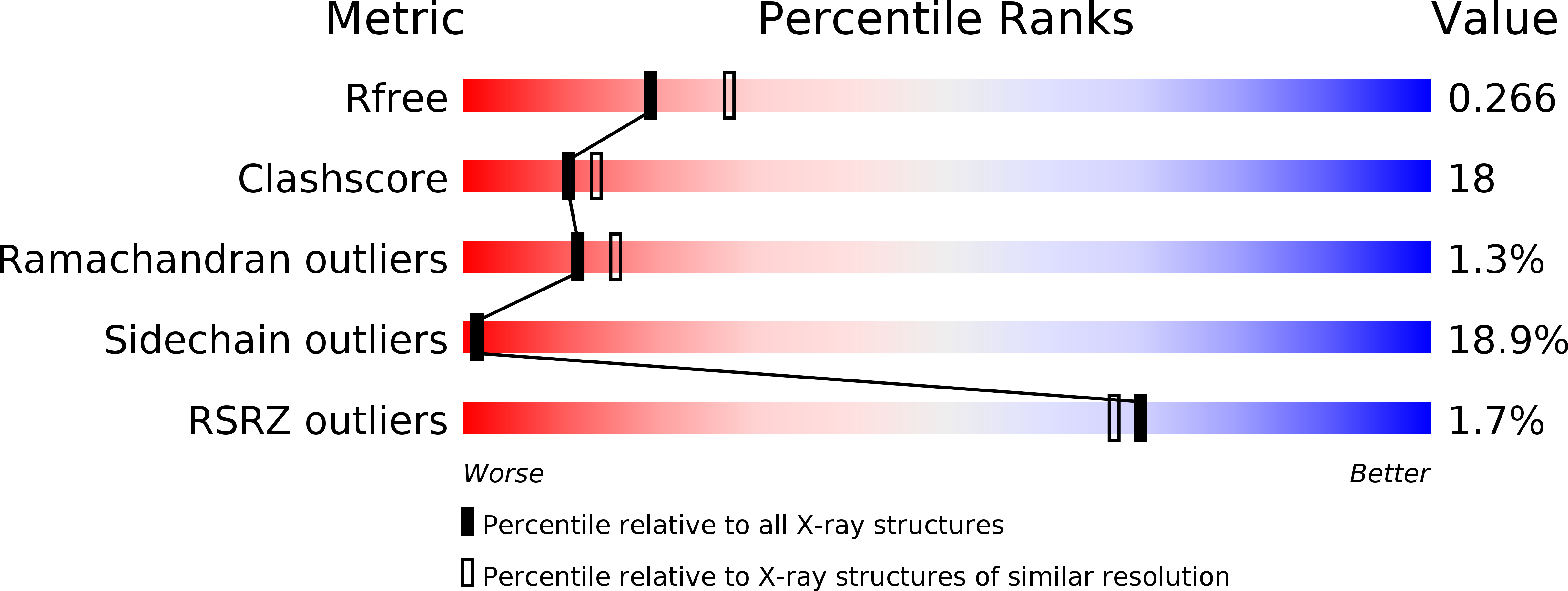
Resolution:
2.42 Å
R-Value Free:
0.25
R-Value Work:
0.22
R-Value Observed:
0.22
Space Group:
P 1 21 1
Macromolecular Entities
Structures with similar UniProt ID
Protein Blast
Polymer Type:polypeptide(L)
Molecule:Lactotransferrin
Gene (Uniprot):LTF
Chain IDs:A, B
Chain Length:348
Number of Molecules:2
Biological Source:Bos taurus
Modified Residue
Compound ID Chain ID Parent Comp ID Details 2D Image
ASN A ASN GLYCOSYLATION SITE
Ligand Molecules
Primary Citation
Structure of the iron-free true C-terminal half of bovine lactoferrin produced by tryptic digestion and its functional significance in the gut.
Febs J. 281 2871 2882 (2014)
PMID: 24798798 DOI: 10.1111/febs.12827

Abstact

Bovine lactoferrin, a 76-kDa glycoprotein (Ala1-Arg689) consists of two similar N- and C-terminal molecular halves with the ability to bind two Fe(3+) ions. The N-terminal half, designated as the N-lobe (Ala1-Arg341) and the C-terminal half designated as the C-lobe (Tyr342-Arg689) have similar iron-binding properties, but the resistant C-lobe prolongs the physiological role of bovine lactoferrin in the digestive tract. Here, we report the crystal structure of true C-lobe, which was produced by limited proteolysis of bovine lactoferrin using trypsin. In the first proteolysis step, two fragments of 21 kDa (Glu86-Lys282) and 45 kDa (Ser283-Arg689) were generated because two lysine residues, Lys85 and Lys282, in the structure of iron-saturated bovine lactoferrin were fully exposed. The 45-kDa fragment was further digested at the newly exposed side chain of Arg341, generating a 38-kDa perfect C-lobe (Tyr342-Arg689). By contrast, the apo-lactoferrin was cut by trypsin only at Arg341, which was exposed in the structure of apo-lactoferrin, whereas the other two sites with Lys85 and Lys282 are inaccessible. The purified iron-saturated C-lobe was crystallized at pH 4.0. The structure was determined by the molecular replacement method using coordinates of the C-terminal half (Arg342-Arg689) of intact camel apo-lactoferrin. The structure determination revealed that the iron atom was absent and the iron-binding cleft was found in a wide-open conformation, whereas in the previously determined structure of iron-saturated C-lobe of bovine lactoferrin, the iron atom was present and the iron-binding site was in the closed confirmation.

Legend

Protein

Chemical

Disease

Primary Citation of related structures