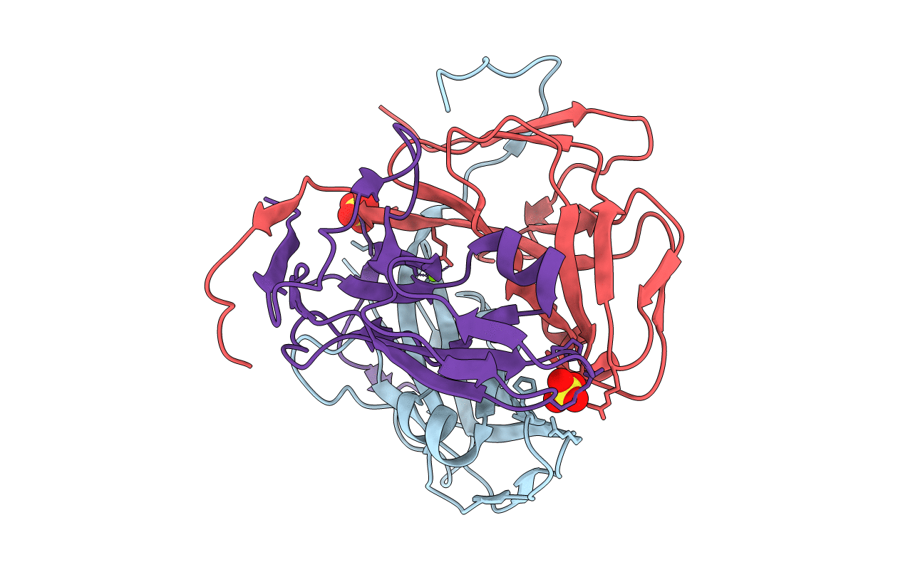
4OOQ image
Deposition Date 2014-02-03
Release Date 2015-07-22
Last Version Date 2023-09-20
Entry Detail
PDB ID:
4OOQ
Keywords:
Title:
apo-dUTPase from Arabidopsis thaliana
Biological Source:
Source Organism:
Host Organism:
Method Details:
Experimental Method:
Resolution:
2.00 Å
R-Value Free:
0.19
R-Value Work:
0.14
R-Value Observed:
0.15
Space Group:
P 21 21 21
Macromolecular Entities
Structures with similar UniProt ID
Protein Blast
Polymer Type:polypeptide(L)
Molecule:Deoxyuridine 5'-triphosphate nucleotidohydrolase
Gene (Uniprot):DUT
Chain IDs:A, B, C
Chain Length:166
Number of Molecules:3
Biological Source:Arabidopsis thaliana
Primary Citation
Structural insights into the mechanism defining substrate affinity in Arabidopsis thaliana dUTPase: the role of tryptophan 93 in ligand orientation.
BMC Res Notes 8 784 784 (2015)
PMID: 26666293 DOI: 10.1186/s13104-015-1760-1

Abstact

BACKGROUND Deoxyuridine triphosphate nucleotidohydrolase (dUTPase) hydrolyzes dUTP to dUMP and pyrophosphate to maintain the cellular thymine-uracil ratio. dUTPase is also a target for cancer chemotherapy. However, the mechanism defining its substrate affinity remains unclear. Sequence comparisons of various dUTPases revealed that Arabidopsis thaliana dUTPase has a unique tryptophan at position 93, which potentially contributes to its degree of substrate affinity. To better understand the roles of tryptophan 93, A. thaliana dUTPase was studied. RESULTS Enzyme assays showed that A. thaliana dUTPase belongs to a high-affinity group of isozymes, which also includes the enzymes from Escherichia coli and Mycobacterium tuberculosis. Enzymes from Homo sapiens and Saccharomyces cerevisiae are grouped as low-affinity dUTPases. The structure of the homo-trimeric A. thaliana dUTPase showed three active sites, each with a different set of ligand interactions between the amino acids and water molecules. On an α-helix, tryptophan 93 appears to keep serine 89 in place via a water molecule and to specifically direct the ligand. Upon being oriented in the active site, the C-terminal residues close the active site to promote the reaction. CONCLUSIONS In the high-affinity group, the prefixed direction of the serine residues was oriented by a positively charged residue located four amino acids away, while low-affinity enzymes possess small hydrophobic residues at the corresponding sites.

Legend

Protein

Chemical

Disease

Primary Citation of related structures