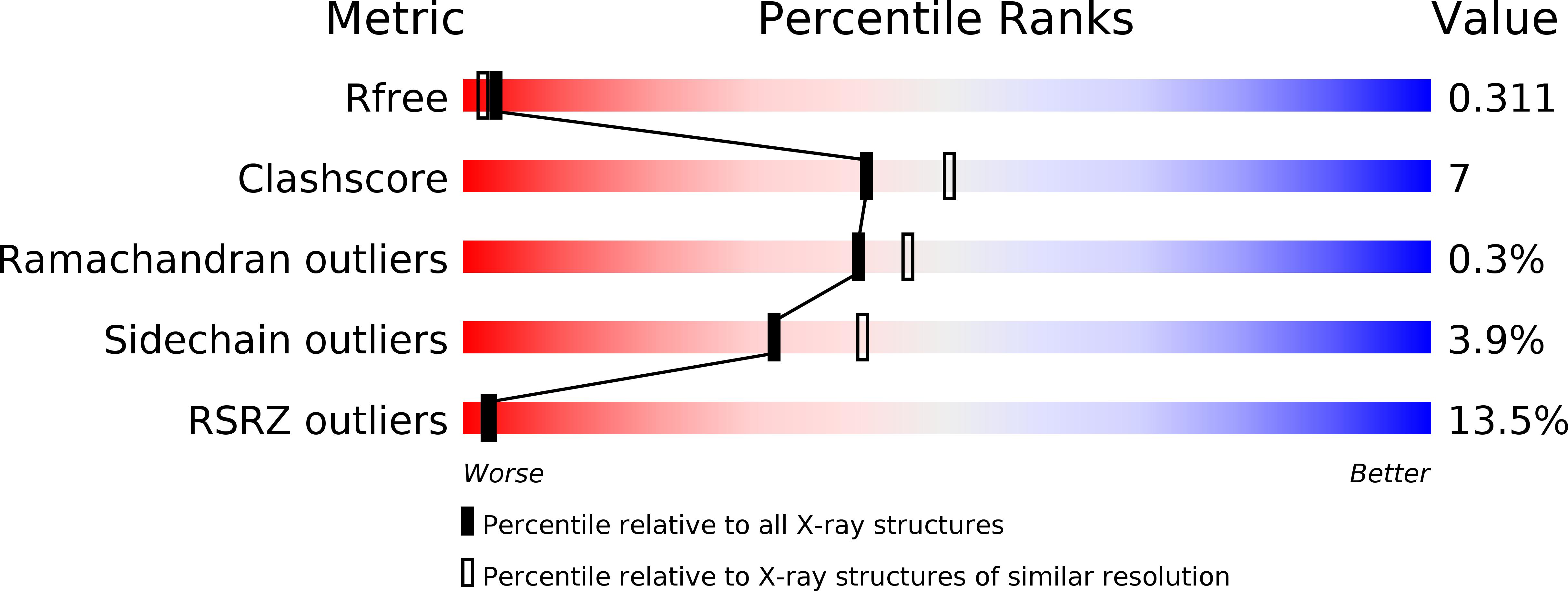
4OOI image
Deposition Date 2014-02-03
Release Date 2014-12-31
Last Version Date 2023-11-08
Entry Detail
PDB ID:
4OOI
Title:
Reduced HlyU from Vibrio cholerae N16961
Biological Source:
Source Organism:
Host Organism:
Method Details:
Experimental Method:
Resolution:
2.20 Å
R-Value Free:
0.30
R-Value Work:
0.25
R-Value Observed:
0.26
Space Group:
C 1 2 1
Macromolecular Entities
Polymer Type:polypeptide(L)
Molecule:Transcriptional activator HlyU
Gene (Uniprot):hlyU
Chain IDs:A, B, C, D
Chain Length:112
Number of Molecules:4
Biological Source:Vibrio cholerae
Primary Citation
Crystal structure of HlyU, the hemolysin gene transcription activator, from Vibrio cholerae N16961 and functional implications
Biochim.Biophys.Acta 1844 2346 2354 (2014)
PMID: 25450504 DOI: 10.1016/j.bbapap.2014.09.020

Abstact

HlyU in Vibrio cholerae is known to be the transcriptional activator of the hemolysin gene, HlyA and possibly a regulator of other virulence factors influencing growth, colonization and pathogenicity of this infective agent. Here we report the crystal structure of HlyU from V. cholerae N16961 (HlyU_Vc) at 1.8Å. The protein, with five α-helices and three β-strands in the topology of α1-α2-β1-α3-α4-β2-β3-α5, forms a homodimer. Helices α3-α4 and a β sheet form the winged helix-turn-helix (wHTH) DNA-binding motif common to the transcription regulators of the SmtB/ArsR family. In spite of an overall fold similar to SmtB/ArsR family, it lacks any metal binding site seen in SmtB. A comparison of the dimeric interfaces showed that the one in SmtB is much larger and have salt bridges that can be disrupted to accommodate metal ions. A model of HlyU-DNA complex suggests bending of the DNA. Cys38 in the structure was found to be modified as sulfenic acid; the oxidized form was not seen in another structure solved under reducing condition. Although devoid of any metal binding site, the presence of a Cys residue exhibiting oxidation-reduction suggests the possibility of the existence of a redox switch in transcription regulation. A structure-based phylogenetic analysis of wHTH proteins revealed the segregation of metal and non-metal binding proteins as well as those in the latter group that are under redox control.

Legend

Protein

Chemical

Disease

Primary Citation of related structures