4OLB image
Deposition Date 2014-01-23
Release Date 2014-02-05
Last Version Date 2023-09-20
Entry Detail
PDB ID:
4OLB
Keywords:
Title:
Crystal Structure of Human Argonaute2 Bound to Tryptophan
Biological Source:
Source Organism:
Homo sapiens (Taxon ID: 9606)
Host Organism:
Method Details:
Experimental Method:
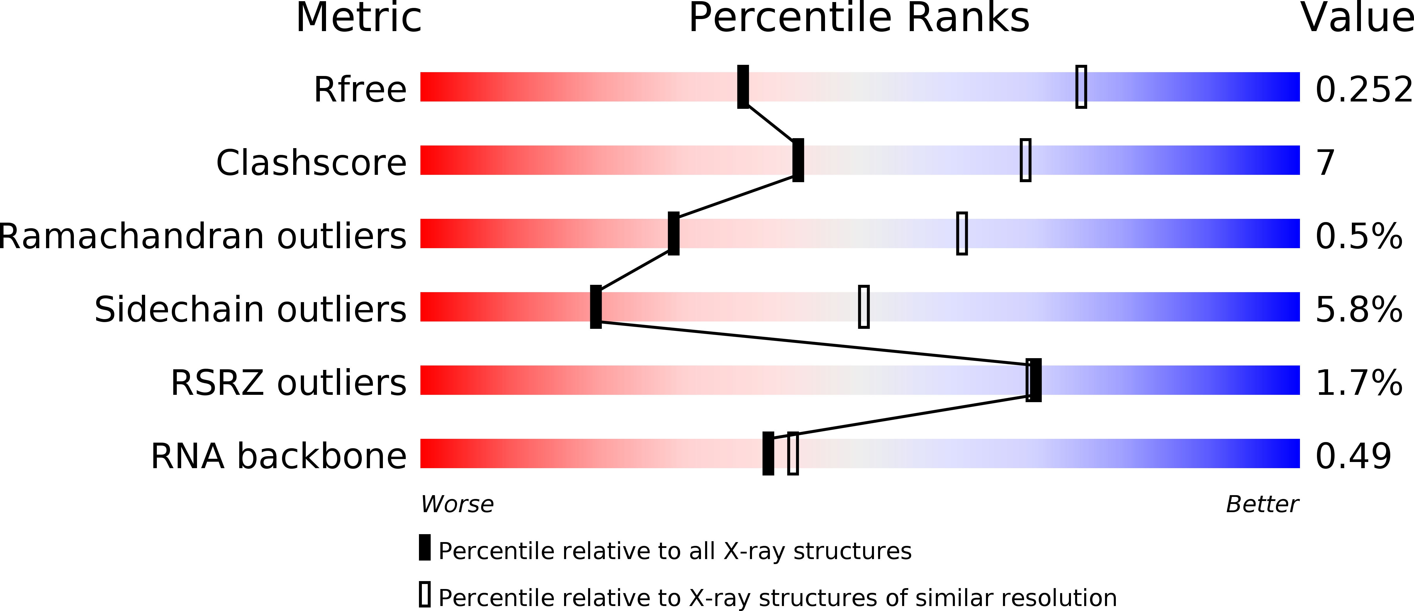
Resolution:
2.90 Å
R-Value Free:
0.24
R-Value Work:
0.20
R-Value Observed:
0.21
Space Group:
P 1 21 1
Macromolecular Entities
Structures with similar UniProt ID
Protein Blast
Polymer Type:polypeptide(L)
Molecule:Protein argonaute-2
Gene (Uniprot):AGO2
Mutagens:S387D
Chain IDs:A
Chain Length:859
Number of Molecules:1
Biological Source:Homo sapiens
Polymer Type:polyribonucleotide
Molecule:5'-R(P*AP*AP*AP*AP*AP*AP*AP*AP*AP*U)-3'
Chain IDs:B
Chain Length:10
Number of Molecules:1
Biological Source:Homo sapiens
Ligand Molecules
Primary Citation
The crystal structure of human Argonaute2.
Science 336 1037 1040 (2012)
PMID: 22539551 DOI: 10.1126/science.1221551

Abstact

Argonaute proteins form the functional core of the RNA-induced silencing complexes that mediate RNA silencing in eukaryotes. The 2.3 angstrom resolution crystal structure of human Argonaute2 (Ago2) reveals a bilobed molecule with a central cleft for binding guide and target RNAs. Nucleotides 2 to 6 of a heterogeneous mixture of guide RNAs are positioned in an A-form conformation for base pairing with target messenger RNAs. Between nucleotides 6 and 7, there is a kink that may function in microRNA target recognition or release of sliced RNA products. Tandem tryptophan-binding pockets in the PIWI domain define a likely interaction surface for recruitment of glycine-tryptophan-182 (GW182) or other tryptophan-rich cofactors. These results will enable structure-based approaches for harnessing the untapped therapeutic potential of RNA silencing in humans.

Legend

Protein

Chemical

Disease

Primary Citation of related structures
Feedback Form
Name
Email
Institute
Feedback