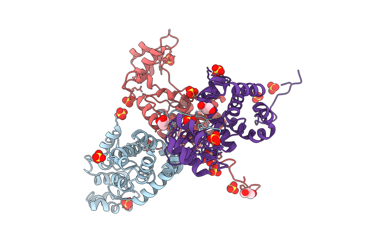
4OK7 image
Deposition Date 2014-01-22
Release Date 2014-03-19
Last Version Date 2024-02-28
Entry Detail
PDB ID:
4OK7
Keywords:
Title:
Structure of bacteriophage SPN1S endolysin from Salmonella typhimurium
Biological Source:
Source Organism:
Host Organism:
Method Details:
Experimental Method:
Resolution:
1.90 Å
R-Value Free:
0.23
R-Value Work:
0.20
Space Group:
P 21 21 21
Macromolecular Entities
Structures with similar UniProt ID
Protein Blast
Polymer Type:polypeptide(L)
Molecule:Endolysin
Gene (Uniprot):SPN1S_0028
Chain IDs:A, B, C
Chain Length:223
Number of Molecules:3
Biological Source:Salmonella phage SPN1S
Primary Citation
Structure of bacteriophage SPN1S endolysin reveals an unusual two-module fold for the peptidoglycan lytic and binding activity.
Mol.Microbiol. 92 316 325 (2014)
PMID: 24641441 DOI: 10.1111/mmi.12555

Abstact

Bacteriophage SPN1S infects the pathogenic Gram-negative bacterium Salmonella typhimurium and expresses endolysin for the release of phage progeny by degrading peptidoglycan of the host cell walls. Bacteriophage SPN1S endolysin exhibits high glycosidase activity against peptidoglycans, resulting in antimicrobial activity against a broad range of outer membrane-permeabilized Gram-negative bacteria. Here, we report a crystal structure of SPN1S endolysin, indicating that unlike most endolysins from Gram-negative bacteria background, the α-helical protein consists of two modular domains, a large and a small domain, with a concave groove between them. Comparison with other structurally homologous glycoside hydrolases indicated a possible peptidoglycan binding site in the groove, and the presence of a catalytic dyad in the vicinity of the groove, one residue in a large domain and the other in a junction between the two domains. The catalytic dyad was further validated by antimicrobial activity assay against outer membrane-permeabilized Escherichia coli. The three-helix bundle in the small domain containing a novel class of sequence motif exhibited binding affinity against outer membrane-permeabilized E. coli and was therefore proposed as the peptidoglycan-binding domain. These structural and functional features suggest that endolysin from a Gram-negative bacterial background has peptidoglycan-binding activity and performs glycoside hydrolase activity through the catalytic dyad.

Legend

Protein

Chemical

Disease

Primary Citation of related structures