4OJ6 image
Entry Detail
PDB ID:
4OJ6
Keywords:
Title:
Crystal Structure of a Putative Tailspike Protein (TSP1, orf210) from Escherichia coli O157:H7 Bacteriohage CBA120; Se-Met Protein
Biological Source:
Source Organism:
Host Organism:
PDB Version:
Deposition Date:
2014-01-20
Release Date:
2014-03-26
Method Details:
Experimental Method:
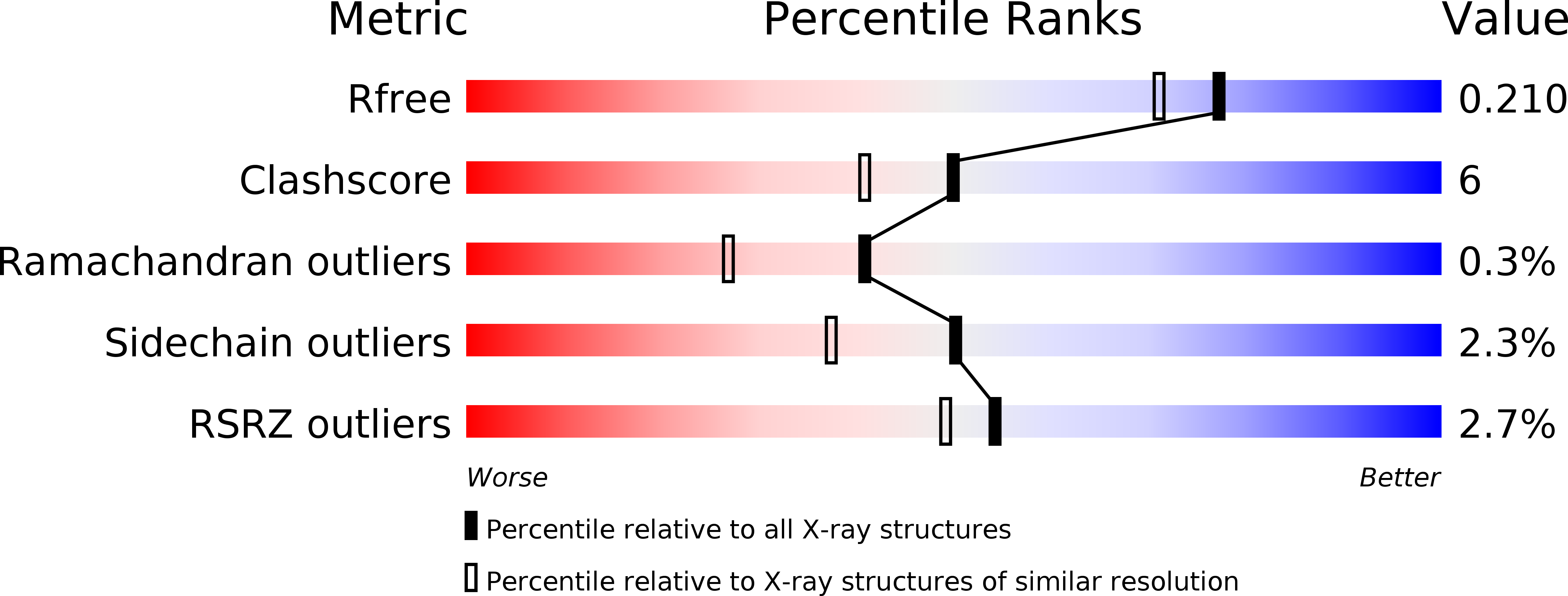
Resolution:
1.80 Å
R-Value Free:
0.20
R-Value Work:
0.18
R-Value Observed:
0.18
Space Group:
P 21 21 21
Macromolecular Entities
Polymer Type:polypeptide(L)
Description:Tailspike protein
Chain IDs:A, B, C
Chain Length:776
Number of Molecules:3
Biological Source:Escherichia phage Cba120
Modified Residue
Compound ID Chain ID Parent Comp ID Details 2D Image
MSE A MET SELENOMETHIONINE
Ligand Molecules
Primary Citation
Crystal structure of ORF210 from E. coli O157:H1 phage CBA120 (TSP1), a putative tailspike protein.
Plos One 9 e93156 e93156 (2014)
PMID: 24671238 DOI: 10.1371/journal.pone.0093156

Abstact

Bacteriophage tailspike proteins act as primary receptors, often possessing endoglycosidase activity toward bacterial lipopolysaccharides or other exopolysaccharides, which enable phage absorption and subsequent DNA injection into the host. Phage CBA120, a contractile long-tailed Viunalikevirus phage infects the virulent Escherichia coli O157:H7. This phage encodes four putative tailspike proteins exhibiting little amino acid sequence identity, whose biological roles and substrate specificities are unknown. Here we focus on the first tailspike, TSP1, encoded by the orf210 gene. We have discovered that TSP1 is resistant to protease degradation, exhibits high thermal stability, but does not cleave the O157 antigen. An immune-dot blot has shown that TSP1 binds strongly to non-O157:H7 E. coli cells and more weakly to K. pneumoniae cells, but exhibits little binding to E. coli O157:H7 strains. To facilitate structure-function studies, we have determined the crystal structure of TSP1 to a resolution limit of 1.8 Å. Similar to other tailspikes proteins, TSP1 assembles into elongated homotrimers. The receptor binding region of each subunit adopts a right-handed parallel β helix, reminiscent yet not identical to several known tailspike structures. The structure of the N-terminal domain that binds to the virion particle has not been seen previously. Potential endoglycosidase catalytic sites at the three subunit interfaces contain two adjacent glutamic acids, unlike any catalytic machinery observed in other tailspikes. To identify potential sugar binding sites, the crystal structures of TSP1 in complexes with glucose, α-maltose, or α-lactose were determined. These structures revealed that each sugar binds in a different location and none of the environments appears consistent with an endoglycosidase catalytic site. Such sites may serve to bind sugar units of a yet to be identified bacterial exopolysaccharide.

Legend

Protein

Chemical

Disease

Primary Citation of related structures