4OHS image
Deposition Date 2014-01-17
Release Date 2014-02-26
Last Version Date 2024-11-27
Entry Detail
PDB ID:
4OHS
Title:
The structure of a far-red fluorescent protein, AQ143
Biological Source:
Source Organism:
Actinia Equina (Taxon ID: 6106)
Method Details:
Experimental Method:
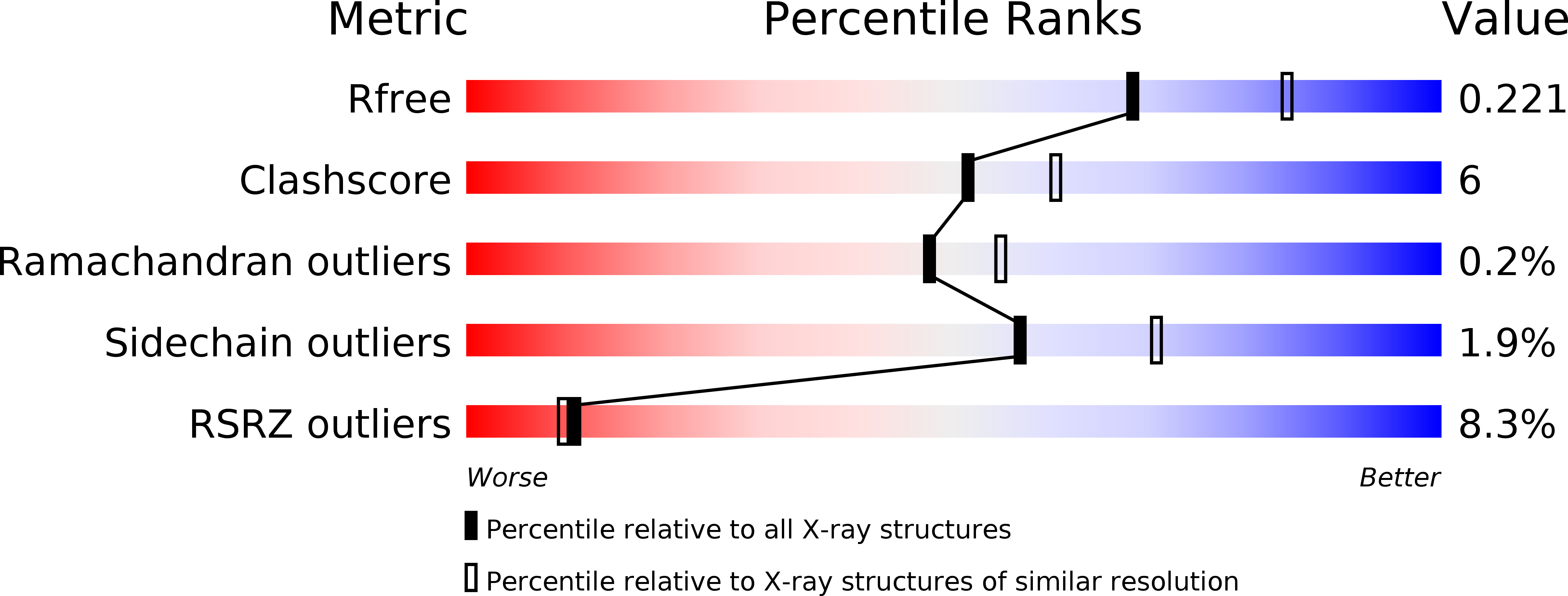
Resolution:
2.19 Å
R-Value Free:
0.22
R-Value Work:
0.18
R-Value Observed:
0.19
Space Group:
P 1
Macromolecular Entities
Structures with similar UniProt ID
Protein Blast
Polymer Type:polypeptide(L)
Molecule:FAR-RED FLUORESCENT PROTEIN AQ143
Chain IDs:A, B, C, D, E, F, G, H
Chain Length:239
Number of Molecules:8
Biological Source:Actinia Equina
Modified Residue
Compound ID Chain ID Parent Comp ID Details 2D Image
CH6 A MET ?
NRQ A MET ?
Ligand Molecules
Primary Citation
The structure of a far-red fluorescent protein, AQ143, shows evidence in support of reported red-shifting chromophore interactions.
Protein Sci. 23 1148 1153 (2014)
PMID: 24888769 DOI: 10.1002/pro.2498

Abstact

Engineering fluorescent proteins (FPs) to emit light at longer wavelengths is a significant focus in the development of the next generation of fluorescent biomarkers, as far-red light penetrates tissue with minimal absorption, allowing better imaging inside of biological hosts. Structure-guided design and directed evolution have led to the discovery of red FPs with significant bathochromic shifts to their emission. Here, we present the crystal structure of one of the most bathochromically shifted FPs reported to date, AQ143, a nine-point mutant of aeCP597, a chromoprotein from Actinia equina. The 2.19 Å resolution structure reveals several important chromophore interactions that contribute to the protein's far-red emission and shows dual occupancy of the green and red chromophores.

Legend

Protein

Chemical

Disease

Primary Citation of related structures
Feedback Form
Name
Email
Institute
Feedback