4OGC image
Deposition Date 2014-01-15
Release Date 2014-02-12
Last Version Date 2024-10-16
Entry Detail
PDB ID:
4OGC
Keywords:
Title:
Crystal structure of the Type II-C Cas9 enzyme from Actinomyces naeslundii
Biological Source:
Source Organism:
Host Organism:
Method Details:
Experimental Method:
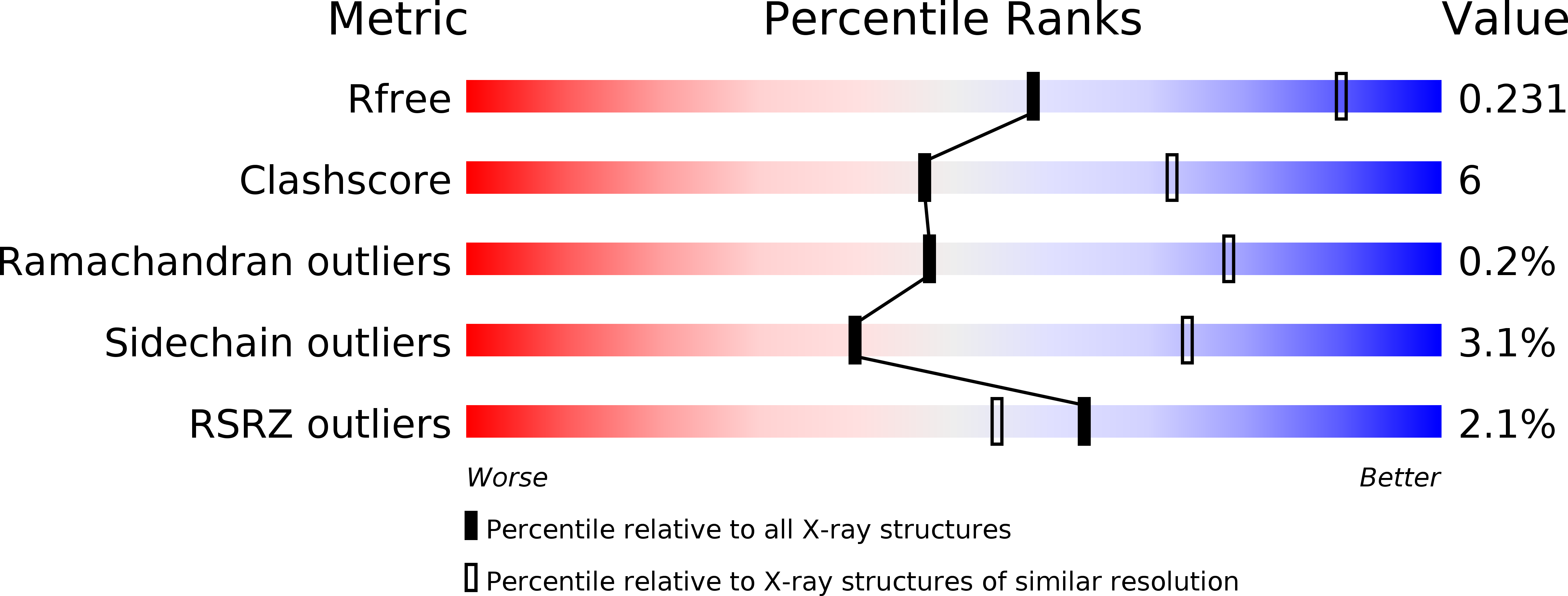
Resolution:
2.80 Å
R-Value Free:
0.23
R-Value Work:
0.19
R-Value Observed:
0.19
Space Group:
P 1 21 1
Macromolecular Entities
Polymer Type:polypeptide(L)
Molecule:HNH endonuclease domain protein
Gene (Uniprot):cas9
Chain IDs:A
Chain Length:1101
Number of Molecules:1
Biological Source:Actinomyces naeslundii
Modified Residue
Compound ID Chain ID Parent Comp ID Details 2D Image
MSE A MET SELENOMETHIONINE
Primary Citation
Structures of Cas9 endonucleases reveal RNA-mediated conformational activation.
Science 343 1247997 1247997 (2014)
PMID: 24505130 DOI: 10.1126/science.1247997

Abstact

Type II CRISPR (clustered regularly interspaced short palindromic repeats)-Cas (CRISPR-associated) systems use an RNA-guided DNA endonuclease, Cas9, to generate double-strand breaks in invasive DNA during an adaptive bacterial immune response. Cas9 has been harnessed as a powerful tool for genome editing and gene regulation in many eukaryotic organisms. We report 2.6 and 2.2 angstrom resolution crystal structures of two major Cas9 enzyme subtypes, revealing the structural core shared by all Cas9 family members. The architectures of Cas9 enzymes define nucleic acid binding clefts, and single-particle electron microscopy reconstructions show that the two structural lobes harboring these clefts undergo guide RNA-induced reorientation to form a central channel where DNA substrates are bound. The observation that extensive structural rearrangements occur before target DNA duplex binding implicates guide RNA loading as a key step in Cas9 activation.

Legend

Protein

Chemical

Disease

Primary Citation of related structures