4OGA image
Entry Detail
PDB ID:
4OGA
Title:
Insulin in complex with Site 1 of the human insulin receptor
Biological Source:
Source Organism:
PDB Version:
Deposition Date:
2014-01-15
Release Date:
2014-08-27
Method Details:
Experimental Method:
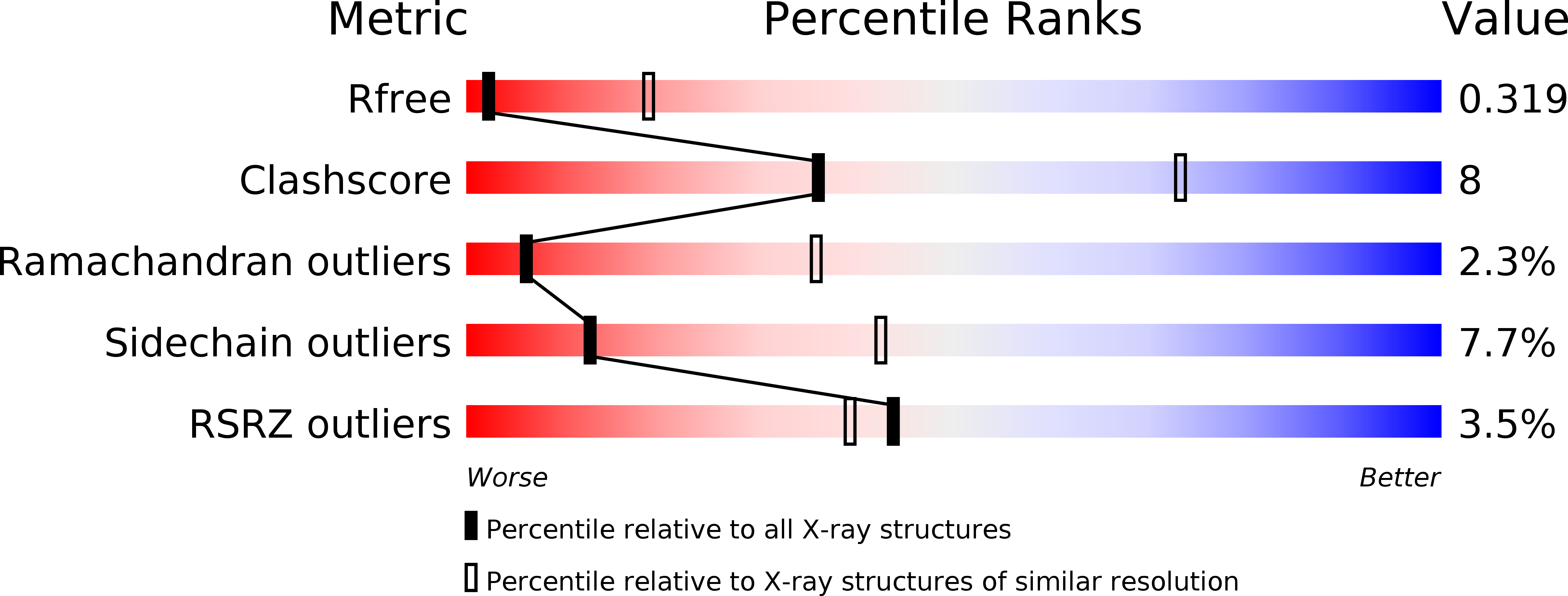
Resolution:
3.50 Å
R-Value Free:
0.28
R-Value Work:
0.26
R-Value Observed:
0.26
Space Group:
P 2 3
Macromolecular Entities
Polymer Type:polypeptide(L)
Description:Insulin A chain
Chain IDs:A
Chain Length:21
Number of Molecules:1
Biological Source:Homo sapiens
Polymer Type:polypeptide(L)
Description:Insulin B chain
Chain IDs:B
Chain Length:30
Number of Molecules:1
Biological Source:Homo sapiens
Polymer Type:polypeptide(L)
Description:monoclonal antibody fab 83-7 fragment - heavy chain
Chain IDs:C
Chain Length:118
Number of Molecules:1
Biological Source:Mus musculus
Polymer Type:polypeptide(L)
Description:monoclonal antibody fab 83-7 fragment - light chain
Chain IDs:D
Chain Length:114
Number of Molecules:1
Biological Source:Mus musculus
Polymer Type:polypeptide(L)
Description:Insulin receptor domains L1-CR
Chain IDs:E
Chain Length:317
Number of Molecules:1
Biological Source:Homo sapiens
Polymer Type:polypeptide(L)
Description:Insulin receptor alpha-CT peptide
Chain IDs:F
Chain Length:16
Number of Molecules:1
Biological Source:Homo sapiens
Modified Residue
Compound ID Chain ID Parent Comp ID Details 2D Image
ASN E ASN GLYCOSYLATION SITE
Ligand Molecules
Primary Citation

Abstact

Insulin provides a classical model of a globular protein, yet how the hormone changes conformation to engage its receptor has long been enigmatic. Interest has focused on the C-terminal B-chain segment, critical for protective self-assembly in β cells and receptor binding at target tissues. Insight may be obtained from truncated "microreceptors" that reconstitute the primary hormone-binding site (α-subunit domains L1 and αCT). We demonstrate that, on microreceptor binding, this segment undergoes concerted hinge-like rotation at its B20-B23 β-turn, coupling reorientation of Phe(B24) to a 60° rotation of the B25-B28 β-strand away from the hormone core to lie antiparallel to the receptor's L1-β2 sheet. Opening of this hinge enables conserved nonpolar side chains (Ile(A2), Val(A3), Val(B12), Phe(B24), and Phe(B25)) to engage the receptor. Restraining the hinge by nonstandard mutagenesis preserves native folding but blocks receptor binding, whereas its engineered opening maintains activity at the price of protein instability and nonnative aggregation. Our findings rationalize properties of clinical mutations in the insulin family and provide a previously unidentified foundation for designing therapeutic analogs. We envisage that a switch between free and receptor-bound conformations of insulin evolved as a solution to conflicting structural determinants of biosynthesis and function.

Legend

Protein

Chemical

Disease

Primary Citation of related structures