4OFC image
Deposition Date 2014-01-14
Release Date 2014-11-19
Last Version Date 2023-09-20
Entry Detail
PDB ID:
4OFC
Keywords:
Title:
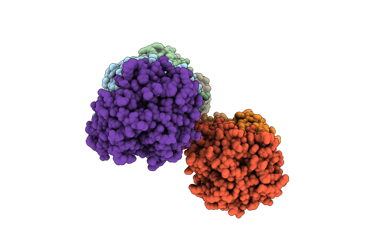
2.0 Angstroms X-ray crystal structure of human 2-amino-3-carboxymuconate-6-semialdehye decarboxylase
Biological Source:
Source Organism:
Homo sapiens (Taxon ID: 9606)
Host Organism:
Method Details:
Experimental Method:
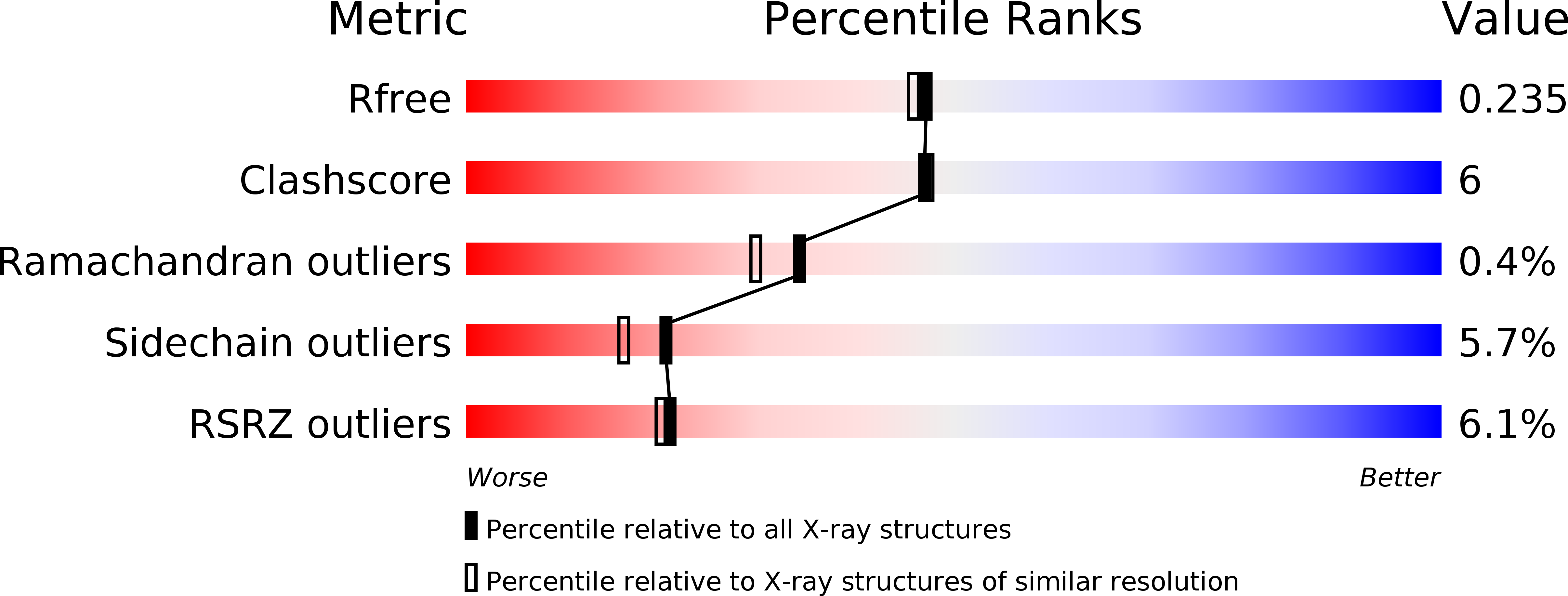
Resolution:
1.99 Å
R-Value Free:
0.24
R-Value Work:
0.19
R-Value Observed:
0.19
Space Group:
P 21 21 21
Macromolecular Entities
Structures with similar UniProt ID
Protein Blast
Polymer Type:polypeptide(L)
Molecule:2-amino-3-carboxymuconate-6-semialdehyde decarboxylase
Gene (Uniprot):ACMSD
Chain IDs:A, B, C, D, E, F
Chain Length:335
Number of Molecules:6
Biological Source:Homo sapiens
Ligand Molecules
Primary Citation
Human alpha-amino-beta-carboxymuconate-epsilon-semialdehyde decarboxylase (ACMSD): A structural and mechanistic unveiling.
Proteins 83 178 187 (2015)
PMID: 25392945 DOI: 10.1002/prot.24722

Abstact

Human α-amino-β-carboxymuconate-ε-semialdehyde decarboxylase determines the fate of tryptophan metabolites in the kynurenine pathway by controlling the quinolinate levels for de novo nicotinamide adenine dinucleotide biosynthesis. The unstable nature of its substrate has made gaining insight into its reaction mechanism difficult. Our electron paramagnetic resonance (EPR) spectroscopic study on the Cu-substituted human enzyme suggests that the native substrate does not directly ligate to the metal ion. Substrate binding did not result in a change of either the hyperfine structure or the super-hyperfine structure of the EPR spectrum. We also determined the crystal structure of the human enzyme in its native catalytically active state (at 1.99 Å resolution), a substrate analogue-bound form (2.50 Å resolution), and a selected active site mutant form with one of the putative substrate binding residues altered (2.32 Å resolution). These structures illustrate that each asymmetric unit contains three pairs of dimers. Consistent with the EPR findings, the ligand-bound complex structure shows that the substrate analogue does not directly coordinate to the metal ion but is bound to the active site by two arginine residues through noncovalent interactions.

Legend

Protein

Chemical

Disease

Primary Citation of related structures