4O96 image
Entry Detail
PDB ID:
4O96
Keywords:
Title:
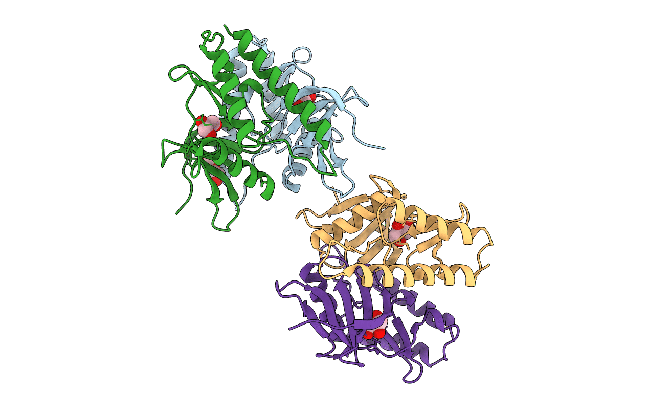
2.60 Angstrom resolution crystal structure of a protein kinase domain of type III effector NleH2 (ECs1814) from Escherichia coli O157:H7 str. Sakai
Biological Source:
Source Organism:
Host Organism:
PDB Version:
Deposition Date:
2014-01-01
Release Date:
2014-01-15
Method Details:
Experimental Method:
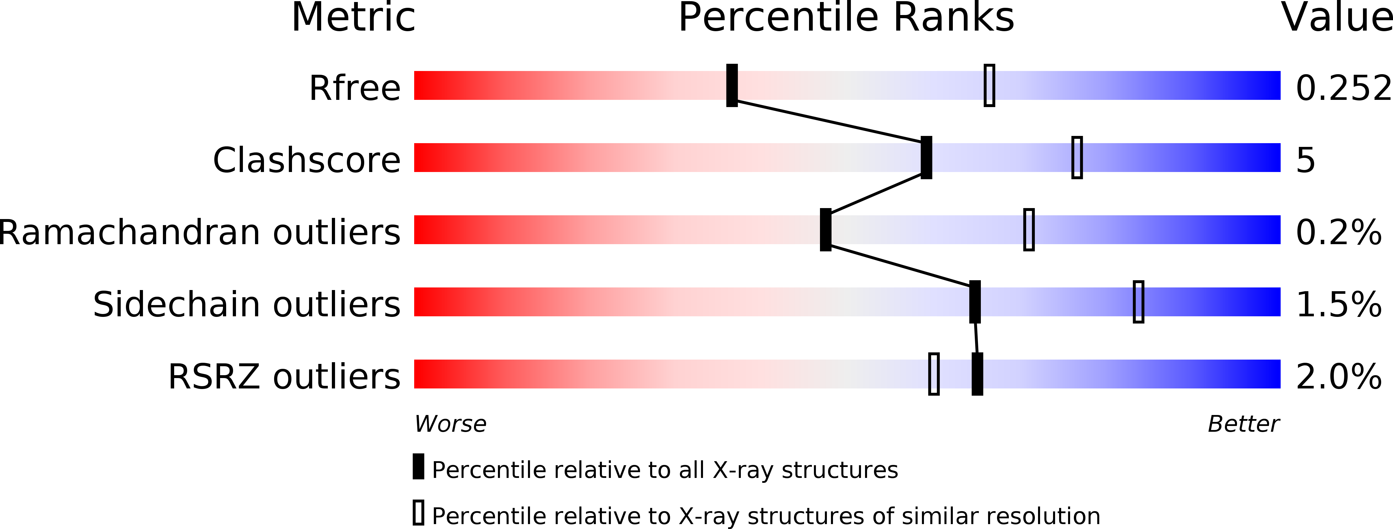
Resolution:
2.60 Å
R-Value Free:
0.25
R-Value Work:
0.20
R-Value Observed:
0.21
Space Group:
P 4 21 2
Macromolecular Entities
Polymer Type:polypeptide(L)
Description:type III effector protein kinase
Mutations:N-terminal 138 residues were truncated
Chain IDs:A, B, C, D
Chain Length:166
Number of Molecules:4
Biological Source:Escherichia coli O157:H7
Primary Citation
Type III Effector NleH2 from Escherichia coli O157:H7 str. Sakai Features an Atypical Protein Kinase Domain.
Biochemistry 53 2433 2435 (2014)
PMID: 24712300 DOI: 10.1021/bi500016j

Abstact

The crystal structure of a C-terminal domain of enterohemorrhagic Escherichia coli type III effector NleH2 has been determined to 2.6 Å resolution. The structure resembles those of protein kinases featuring the catalytic, activation, and glycine-rich loop motifs and ATP-binding site. The position of helix αC and the lack of a conserved arginine within an equivalent HRD motif suggested that the NleH2 kinase domain's active conformation might not require phosphorylation. The activation segment markedly contributed to the dimerization interface of NleH2, which can also accommodate the NleH1-NleH2 heterodimer. The C-terminal PDZ-binding motif of NleH2 provided bases for interaction with host proteins.

Legend

Protein

Chemical

Disease

Primary Citation of related structures