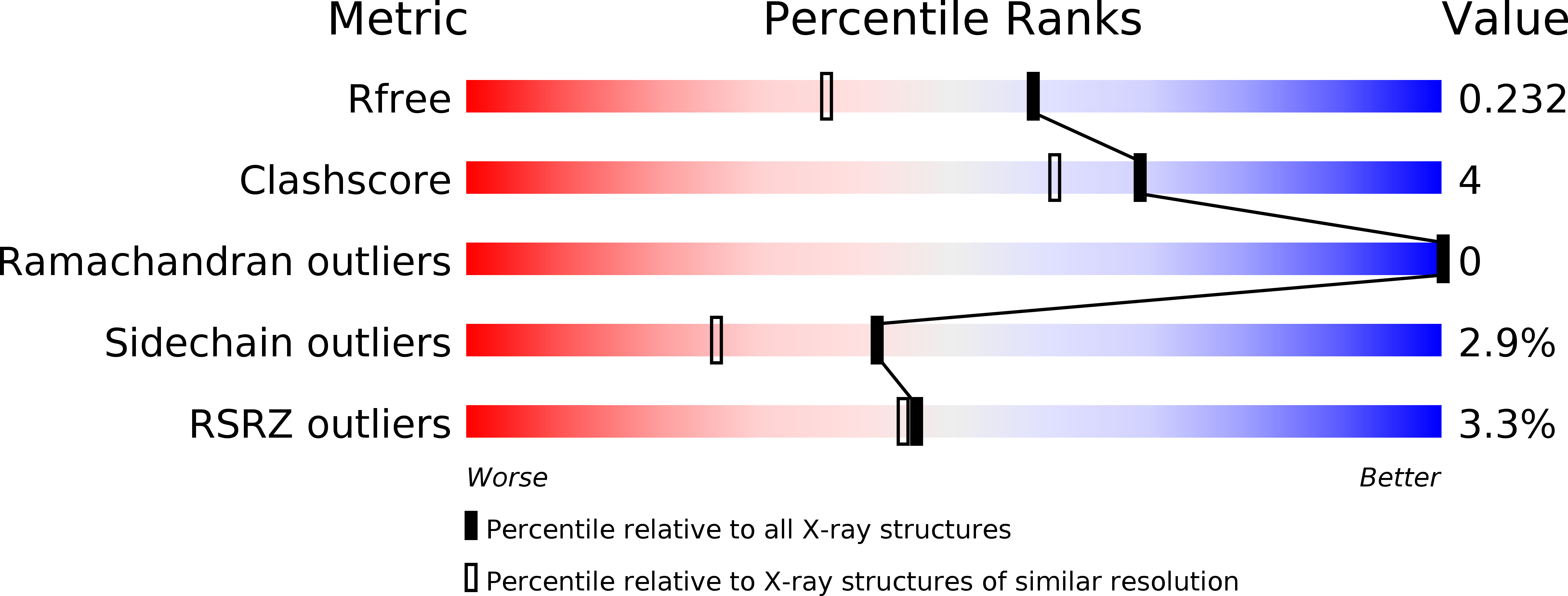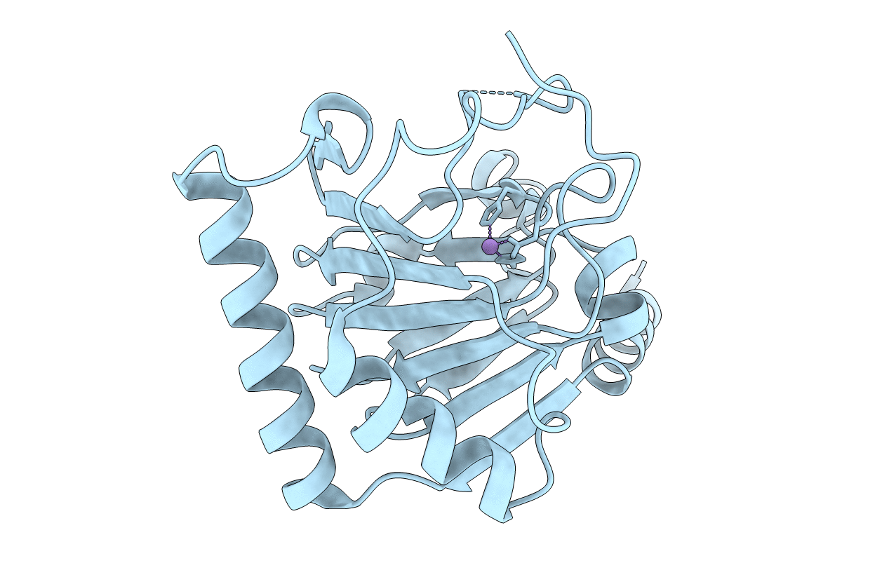
4O7X image
Deposition Date 2013-12-26
Release Date 2014-03-19
Last Version Date 2024-10-09
Entry Detail
PDB ID:
4O7X
Keywords:
Title:
Crystal structure of human ALKBH5 in complex with Mn2+
Biological Source:
Source Organism:
Homo sapiens (Taxon ID: 9606)
Host Organism:
Method Details:
Experimental Method:
Resolution:
1.78 Å
R-Value Free:
0.23
R-Value Work:
0.21
R-Value Observed:
0.21
Space Group:
P 43 21 2
Macromolecular Entities
Structures with similar UniProt ID
Protein Blast
Polymer Type:polypeptide(L)
Molecule:RNA demethylase ALKBH5
Gene (Uniprot):ALKBH5
Chain IDs:A
Chain Length:230
Number of Molecules:1
Biological Source:Homo sapiens
Ligand Molecules
Primary Citation
Crystal structures of the human RNA demethylase Alkbh5 reveal basis for substrate recognition
J.Biol.Chem. 289 11571 11583 (2014)
PMID: 24616105 DOI: 10.1074/jbc.M113.546168

Abstact

N(6)-Methylation of adenosine is the most ubiquitous and abundant modification of nucleoside in eukaryotic mRNA and long non-coding RNA. This modification plays an essential role in the regulation of mRNA translation and RNA metabolism. Recently, human AlkB homolog 5 (Alkbh5) and fat mass- and obesity-associated protein (FTO) were shown to erase this methyl modification on mRNA. Here, we report five high resolution crystal structures of the catalytic core of Alkbh5 in complex with different ligands. Compared with other AlkB proteins, Alkbh5 displays several unique structural features on top of the conserved double-stranded β-helix fold typical of this protein family. Among the unique features, a distinct "lid" region of Alkbh5 plays a vital role in substrate recognition and catalysis. An unexpected disulfide bond between Cys-230 and Cys-267 is crucial for the selective binding of Alkbh5 to single-stranded RNA/DNA by bringing a "flipping" motif toward the central β-helix fold. We generated a substrate binding model of Alkbh5 based on a demethylation activity assay of several structure-guided site-directed mutants. Crystallographic and biochemical studies using various analogs of α-ketoglutarate revealed that the active site cavity of Alkbh5 is much smaller than that of FTO and preferentially binds small molecule inhibitors. Taken together, our findings provide a structural basis for understanding the substrate recognition specificity of Alkbh5 and offer a foundation for selective drug design against AlkB members.

Legend

Protein

Chemical

Disease

Primary Citation of related structures