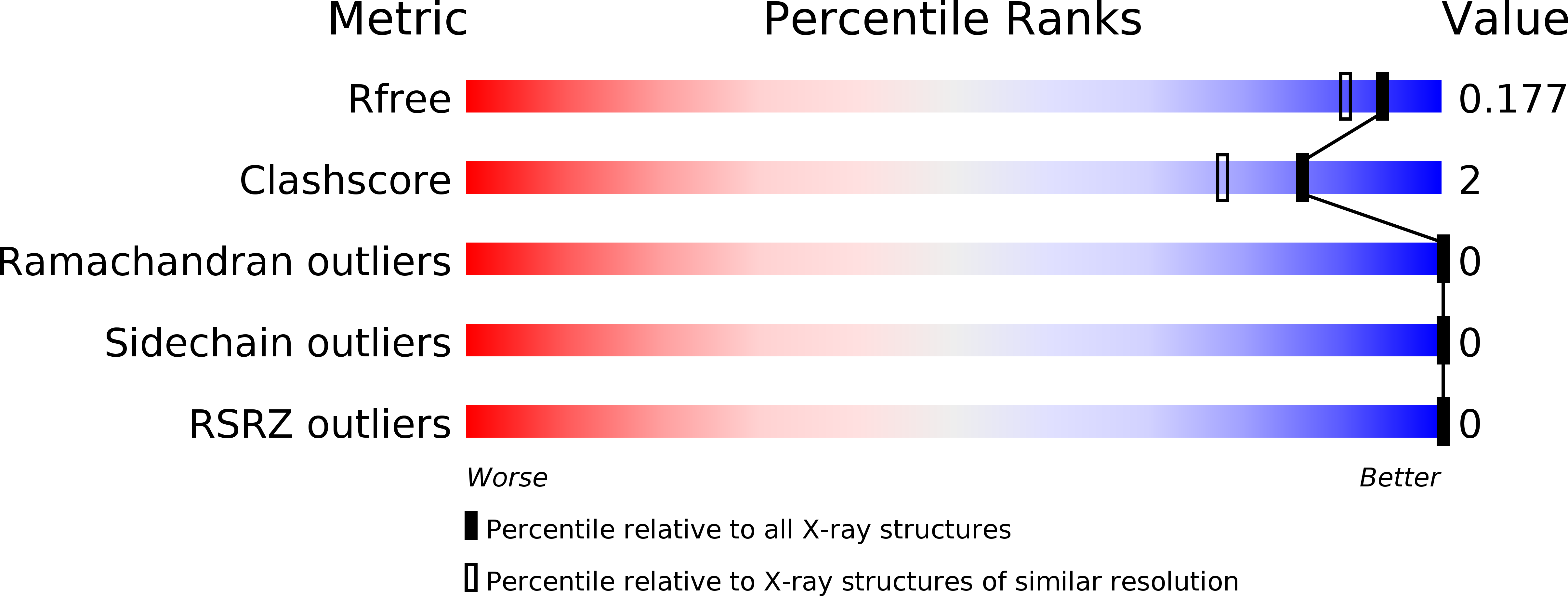
4O6G image
Deposition Date 2013-12-20
Release Date 2014-03-05
Last Version Date 2024-04-03
Entry Detail
PDB ID:
4O6G
Title:
Rv3902c from M. tuberculosis
Biological Source:
Source Organism:
Host Organism:
Method Details:
Experimental Method:
Resolution:
1.55 Å
R-Value Free:
0.17
R-Value Work:
0.16
R-Value Observed:
0.16
Space Group:
P 61
Macromolecular Entities
Polymer Type:polypeptide(L)
Molecule:Uncharacterized protein
Gene (Uniprot):MT4020
Chain IDs:A
Chain Length:174
Number of Molecules:1
Biological Source:Mycobacterium tuberculosis
Primary Citation
1.55 angstrom resolution X-ray crystal structure of Rv3902c from Mycobacterium tuberculosis.
Acta Crystallogr F Struct Biol Commun 70 414 417 (2014)
PMID: 24699730 DOI: 10.1107/S2053230X14003793

Abstact

The crystallographic structure of the Mycobacterium tuberculosis (TB) protein Rv3902c (176 residues; molecular mass of 19.8 kDa) was determined at 1.55 Å resolution. The function of Rv3902c is unknown, although several TB genes involved in bacterial pathogenesis are expressed from the operon containing the Rv3902c gene. The unique structural fold of Rv3902c contains two domains, each consisting of antiparallel β-sheets and α-helices, creating a hand-like binding motif with a small binding pocket in the palm. Structural homology searches reveal that Rv3902c has an overall structure similar to that of the Salmonella virulence-factor chaperone InvB, with an r.m.s.d. for main-chain atoms of 2.3 Å along an aligned domain.

Legend

Protein

Chemical

Disease

Primary Citation of related structures