4O65 image
Deposition Date 2013-12-20
Release Date 2014-04-02
Last Version Date 2024-10-16
Entry Detail
PDB ID:
4O65
Keywords:
Title:
Crystal structure of the cupredoxin domain of amoB from Nitrosocaldus yellowstonii
Biological Source:
Source Organism:
Host Organism:
Method Details:
Experimental Method:
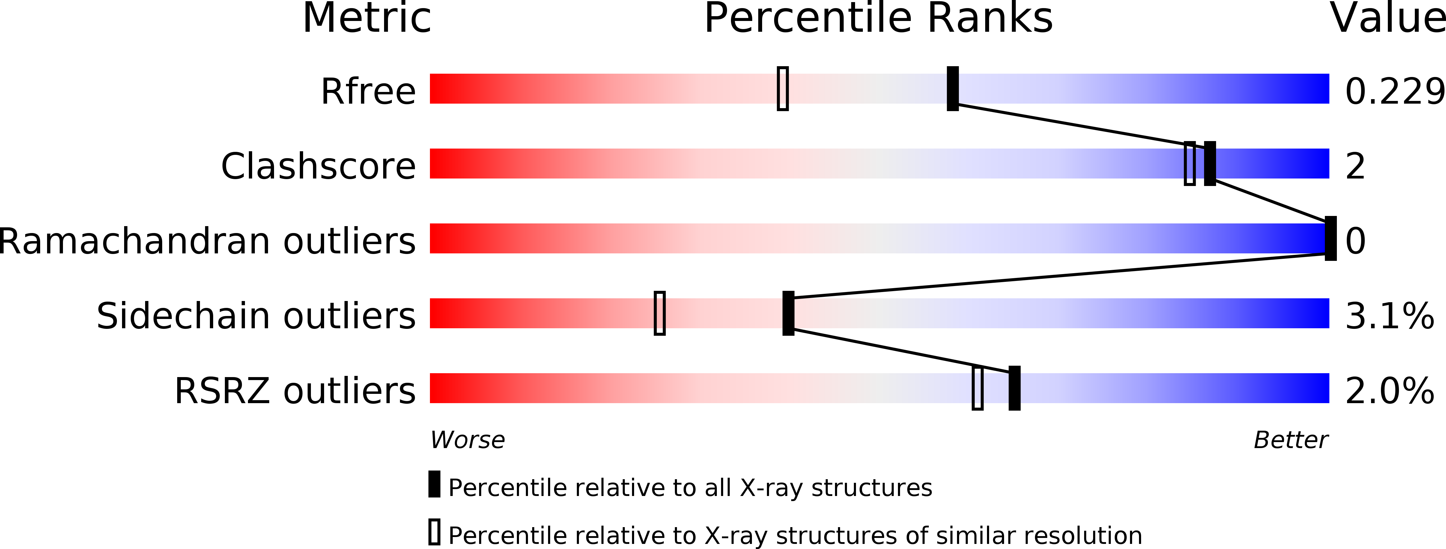
Resolution:
1.80 Å
R-Value Free:
0.22
R-Value Work:
0.18
R-Value Observed:
0.18
Space Group:
P 43 21 2
Macromolecular Entities
Structures with similar UniProt ID
Protein Blast
Polymer Type:polypeptide(L)
Molecule:Putative archaeal ammonia monooxygenase subunit B
Gene (Uniprot):amoB
Chain IDs:A
Chain Length:166
Number of Molecules:1
Biological Source:Candidatus Nitrosocaldus yellowstonii
Primary Citation
Structural conservation of the B subunit in the ammonia monooxygenase/particulate methane monooxygenase superfamily.
Proteins 82 2263 2267 (2014)
PMID: 24523098 DOI: 10.1002/prot.24535

Abstact

The ammonia monooxygenase (AMO)/particulate methane monooxygenase (pMMO) superfamily is a diverse group of membrane-bound enzymes of which only pMMO has been characterized on the molecular level. The pMMO active site is believed to reside in the soluble N-terminal region of the pmoB subunit. To understand the degree of structural conservation within this superfamily, the crystal structure of the corresponding domain of an archaeal amoB subunit from Nitrosocaldus yellowstonii has been determined to 1.8 Å resolution. The structure reveals a remarkable conservation of overall fold and copper binding site location as well as several notable differences that may have implications for function and stability.

Legend

Protein

Chemical

Disease

Primary Citation of related structures