Deposition Date
2013-12-17
Release Date
2014-03-26
Last Version Date
2024-02-28
Entry Detail
PDB ID:
4O30
Keywords:
Title:
Crystal structure of ATXR5 in complex with histone H3.1 and AdoHcy
Biological Source:
Source Organism(s):
Ricinus communis (Taxon ID: 3988)
Expression System(s):
Method Details:
Experimental Method:
Resolution:
2.10 Å
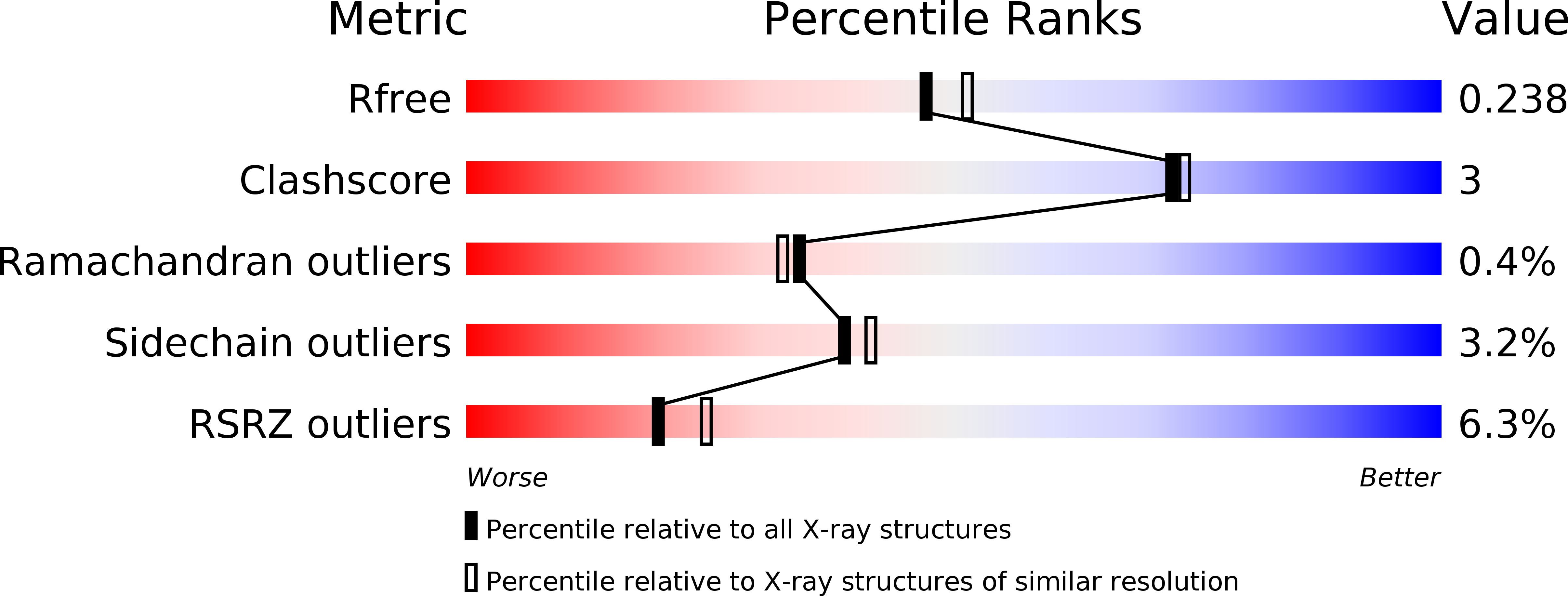
R-Value Free:
0.24
R-Value Work:
0.20
R-Value Observed:
0.20
Space Group:
C 1 2 1