4O1P image
Entry Detail
PDB ID:
4O1P
Title:
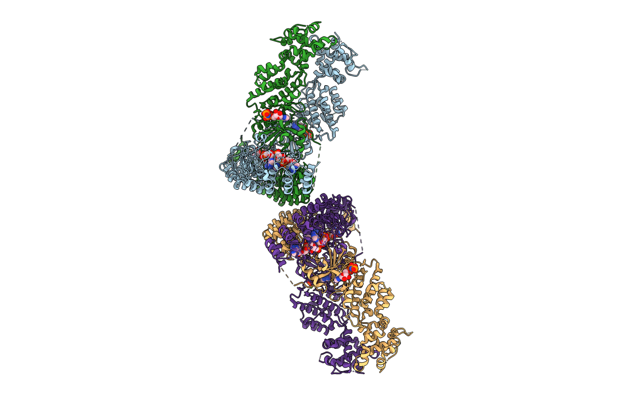
Crystal Structure of RNase L in complex with 2-5A and AMP-PNP
Biological Source:
Source Organism:
Host Organism:
PDB Version:
Deposition Date:
2013-12-16
Release Date:
2014-02-05
Method Details:
Experimental Method:
Resolution:
2.50 Å
R-Value Free:
0.23
R-Value Work:
0.19
R-Value Observed:
0.19
Space Group:
P 1 21 1
Macromolecular Entities
Polymer Type:polypeptide(L)
Description:Ribonuclease L
Chain IDs:A, B, C, D
Chain Length:717
Number of Molecules:4
Biological Source:Sus scrofa
Primary Citation
Dimeric structure of pseudokinase RNase L bound to 2-5A reveals a basis for interferon-induced antiviral activity.
Mol.Cell 53 221 234 (2014)
PMID: 24462203 DOI: 10.1016/j.molcel.2013.12.025

Abstact

RNase L is an ankyrin repeat domain-containing dual endoribonuclease-pseudokinase that is activated by unusual 2,'5'-oligoadenylate (2-5A) second messengers and which impedes viral infections in higher vertebrates. Despite its importance in interferon-regulated antiviral innate immunity, relatively little is known about its precise mechanism of action. Here we present a functional characterization of 2.5 Å and 3.25 Å X-ray crystal and small-angle X-ray scattering structures of RNase L bound to a natural 2-5A activator with and without ADP or the nonhydrolysable ATP mimetic AMP-PNP. These studies reveal how recognition of 2-5A through interactions with the ankyrin repeat domain and the pseudokinase domain, together with nucleotide binding, imposes a rigid intertwined dimer configuration that is essential for RNase catalytic and antiviral functions. The involvement of the pseudokinase domain of RNase L in 2-5A sensing, nucleotide binding, dimerization, and ribonuclease functions highlights the evolutionary adaptability of the eukaryotic protein kinase fold.

Legend

Protein

Chemical

Disease

Primary Citation of related structures