Deposition Date
2013-12-12
Release Date
2014-01-01
Last Version Date
2024-10-30
Entry Detail
PDB ID:
4NZC
Keywords:
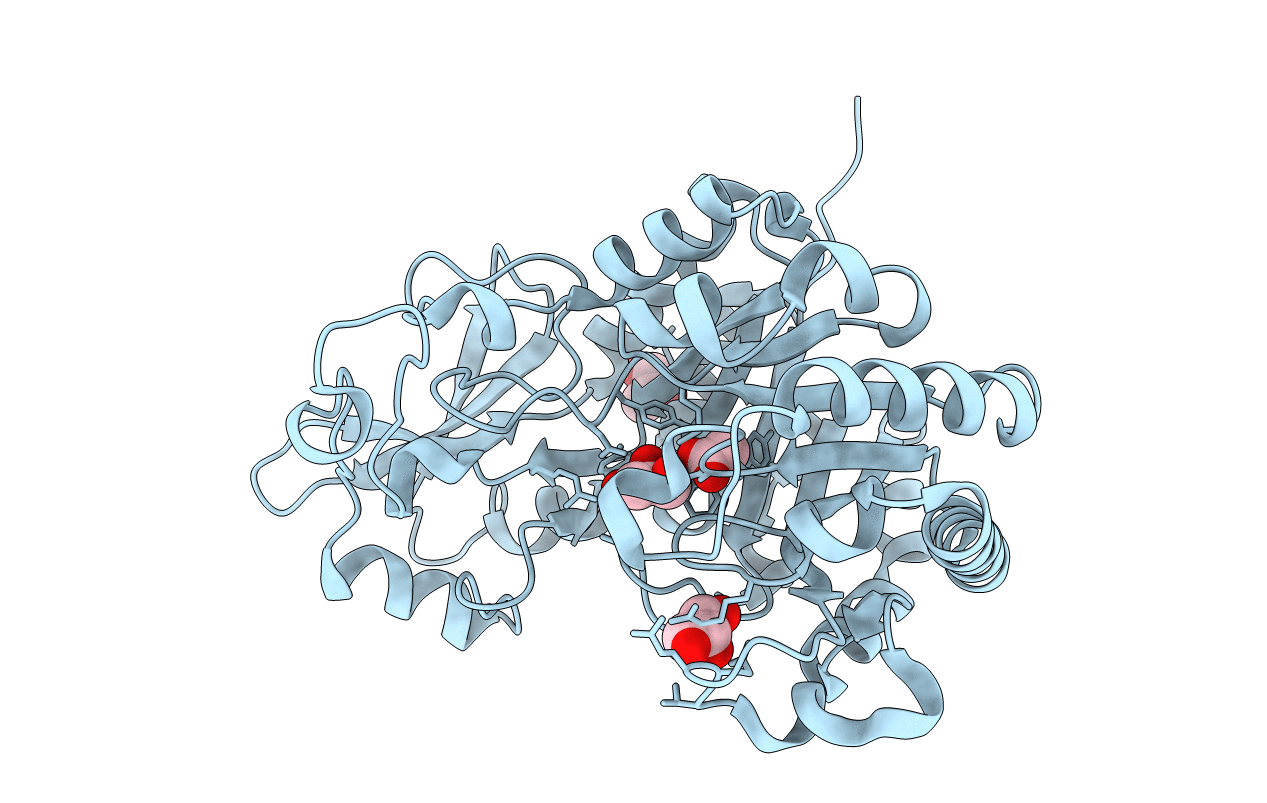
Title:
Crystal structure of Chitinase D from Serratia proteamaculans at 1.45 Angstrom resolution
Biological Source:
Source Organism(s):
Serratia proteamaculans (Taxon ID: 399741)
Expression System(s):
Method Details:
Experimental Method:
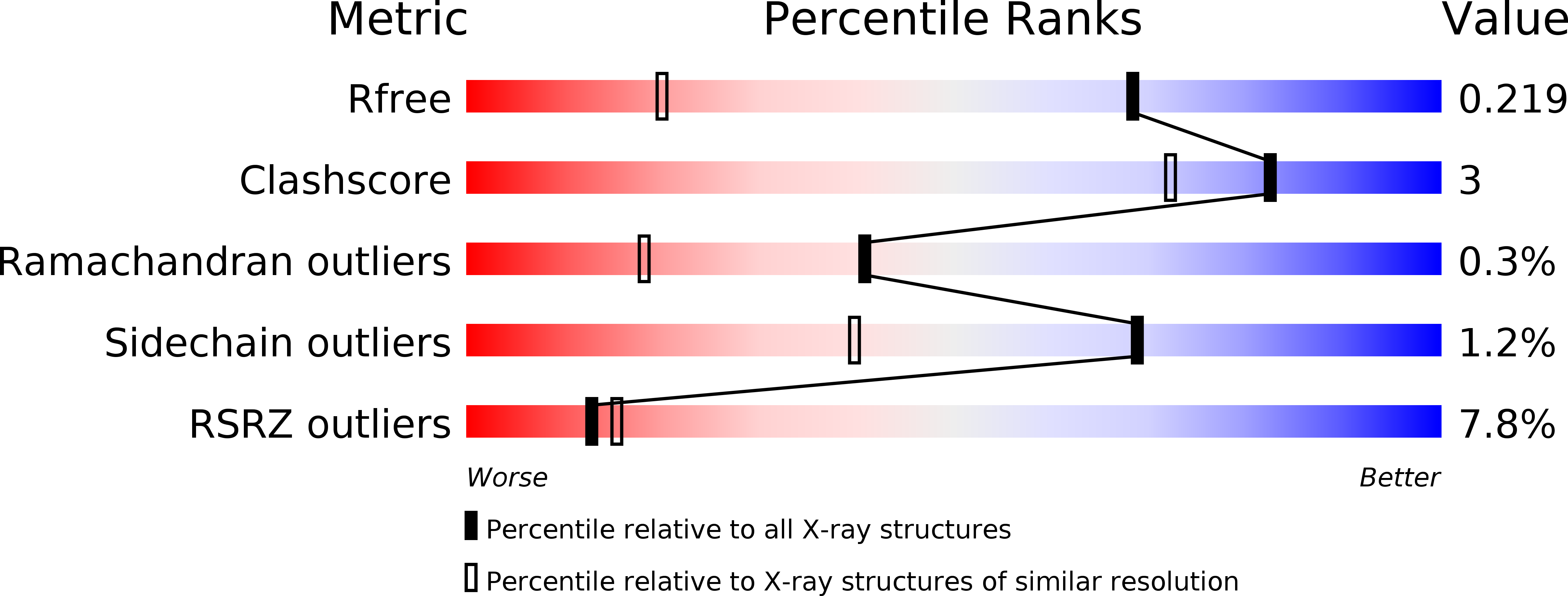
Resolution:
1.45 Å
R-Value Free:
0.21
R-Value Work:
0.19
R-Value Observed:
0.20
Space Group:
P 2 21 21