4NY5 image
Deposition Date 2013-12-10
Release Date 2014-03-26
Last Version Date 2024-11-27
Entry Detail
PDB ID:
4NY5
Keywords:
Title:
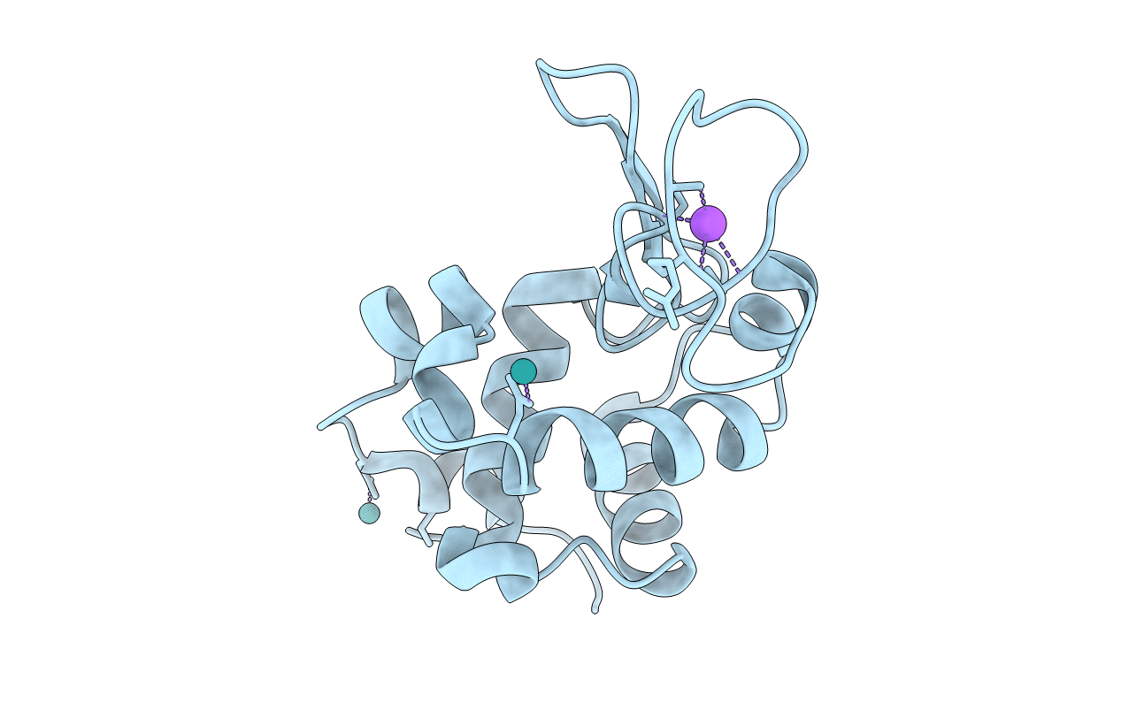
X-ray structure of the adduct formed between hen egg white lysozyme and NAMI-A
Biological Source:
Source Organism:
Gallus gallus (Taxon ID: 9031)
Method Details:
Experimental Method:
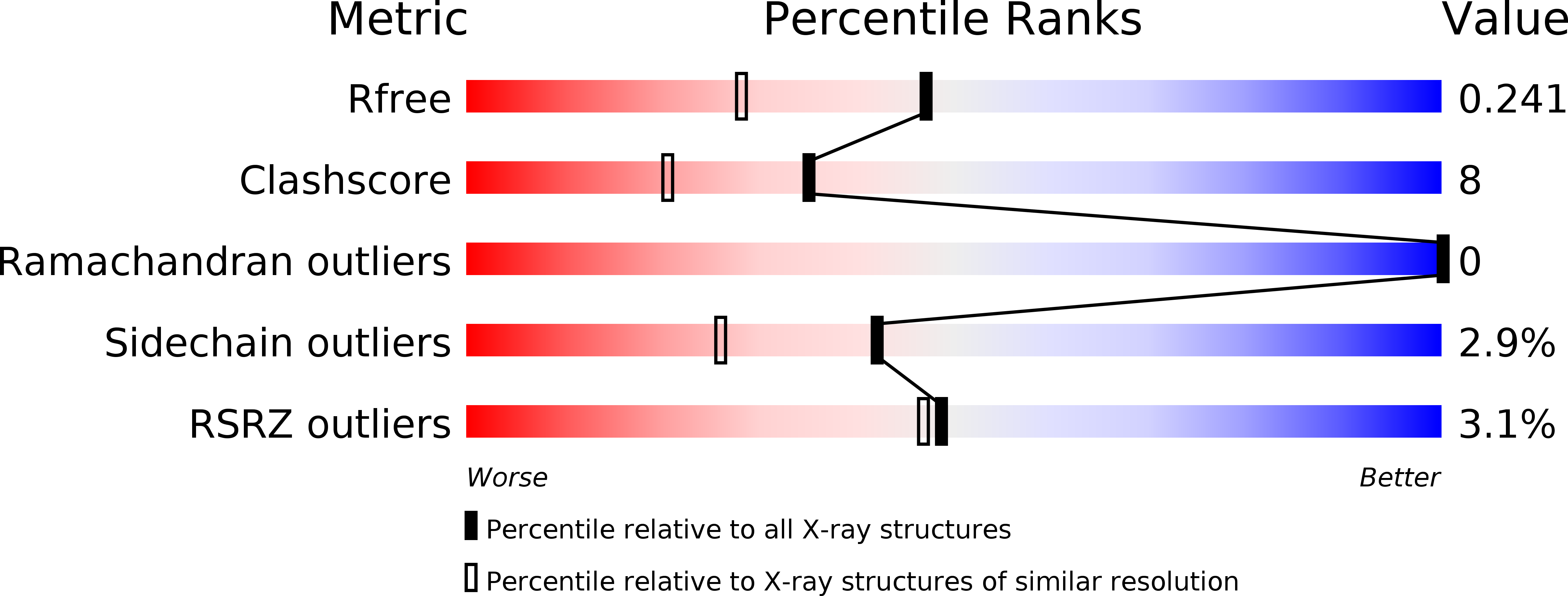
Resolution:
1.85 Å
R-Value Free:
0.23
R-Value Work:
0.17
R-Value Observed:
0.17
Space Group:
P 43 21 2
Macromolecular Entities
Structures with similar UniProt ID
Protein Blast
Polymer Type:polypeptide(L)
Molecule:Lysozyme C
Gene (Uniprot):LYZ
Chain IDs:A
Chain Length:129
Number of Molecules:1
Biological Source:Gallus gallus
Primary Citation
Ruthenium metalation of proteins: the X-ray structure of the complex formed between NAMI-A and hen egg white lysozyme.
Dalton Trans 43 6128 6131 (2014)
PMID: 24553967 DOI: 10.1039/c3dt53582g

Abstact

A crystallographic study of the adduct formed between hen egg white lysozyme (HEWL) and NAMI-A, an established ruthenium(III) anticancer agent in clinical trials, is presented here. The X-ray structure reveals that NAMI-A coordinates the protein, as a naked ruthenium ion, at two distinct sites (namely Asp101 or Asp119) after releasing all its original ligands (DMSO, imidazole and Cl(-)). Structural data of the HEWL/NAMI-A adduct are compared with those previously obtained for the HEWL adduct of AziRu, a NAMI-A analogue bearing a pyridine in place of imidazole. The present results further support the view that NAMI-A exerts its biological effects acting as a classical "prodrug" first undergoing activation and then causing extensive metalation of relevant protein targets. It is also proposed that the original Ru-ligands, although absent in the final adduct, play a major role in directing the ruthenium center to its ultimate anchoring site on the protein surface.

Legend

Protein

Chemical

Disease

Primary Citation of related structures
Feedback Form
Name
Email
Institute
Feedback