4NV1 image
Deposition Date 2013-12-04
Release Date 2013-12-25
Last Version Date 2024-02-28
Entry Detail
PDB ID:
4NV1
Keywords:
Title:
Crystal structure of a 4-N formyltransferase from Francisella tularensis
Biological Source:
Host Organism:
Method Details:
Experimental Method:
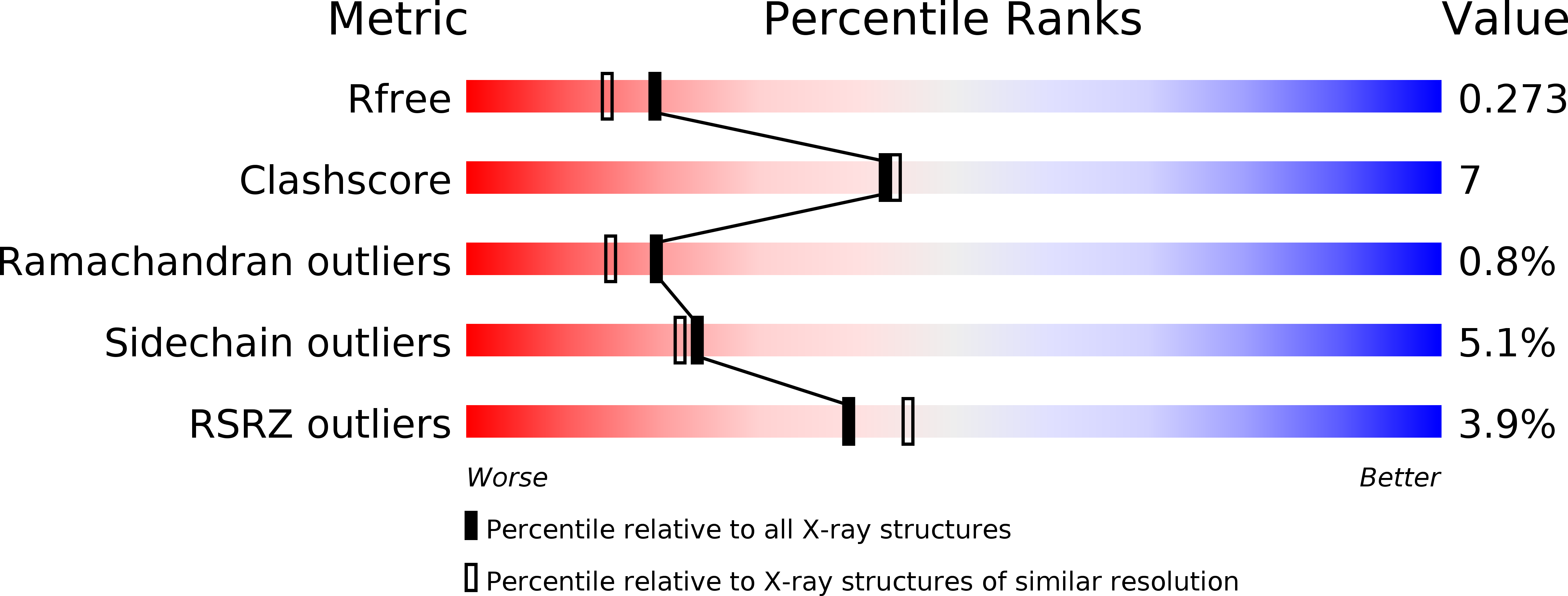
Resolution:
2.10 Å
R-Value Free:
0.27
R-Value Work:
0.19
R-Value Observed:
0.19
Space Group:
P 1
Macromolecular Entities
Structures with similar UniProt ID
Protein Blast
Polymer Type:polypeptide(L)
Molecule:Formyltransferase
Chain IDs:A (auth: C), B (auth: D), C (auth: E), D (auth: F), E (auth: B), F (auth: A), G, H
Chain Length:243
Number of Molecules:8
Biological Source:Francisella tularensis subsp. tularensis
Primary Citation
Three-dimensional structure of a sugar N-formyltransferase from Francisella tularensis.
Protein Sci. 23 273 283 (2014)
PMID: 24347283 DOI: 10.1002/pro.2409

Abstact

N-formylated sugars have been observed on the O-antigens of such pathogenic Gram-negative bacteria as Campylobacter jejuni and Francisella tularensis. Until recently, however, little was known regarding the overall molecular architectures of the N-formyltransferases that are required for the biosynthesis of these unusual sugars. Here we demonstrate that the protein encoded by the wbtj gene from F. tularensis is an N-formyltransferase that functions on dTDP-4-amino-4,6-dideoxy-d-glucose as its substrate. The enzyme, hereafter referred to as WbtJ, demonstrates a strict requirement for N(10) -formyltetrahydrofolate as its carbon source. In addition to the kinetic analysis, the three-dimensional structure of the enzyme was solved in the presence of dTDP-sugar ligands to a nominal resolution of 2.1 Å. Each subunit of the dimeric enzyme is dominated by a "core" domain defined by Met 1 to Ser 185. This core motif harbors the active site residues. Following the core domain, the last 56 residues fold into two α-helices and a β-hairpin motif. The hairpin motif is responsible primarily for the subunit:subunit interface, which is characterized by a rather hydrophobic pocket. From the study presented here, it is now known that WbtJ functions on C-4' amino sugars. Another enzyme recently investigated in the laboratory, WlaRD, formylates only C-3' amino sugars. Strikingly, the quaternary structures of WbtJ and WlaRD are remarkably different. In addition, there are several significant variations in the side chains that line their active site pockets, which may be important for substrate specificity. Details concerning the kinetic and structural properties of WbtJ are presented.

Legend

Protein

Chemical

Disease

Primary Citation of related structures
Feedback Form
Name
Email
Institute
Feedback