4NUB image
Deposition Date 2013-12-03
Release Date 2014-02-26
Last Version Date 2024-10-30
Entry Detail
PDB ID:
4NUB
Keywords:
Title:
Crystal structure of Escherichia coli ribosomal oxygenase YcfD
Biological Source:
Source Organism:
Host Organism:
Method Details:
Experimental Method:
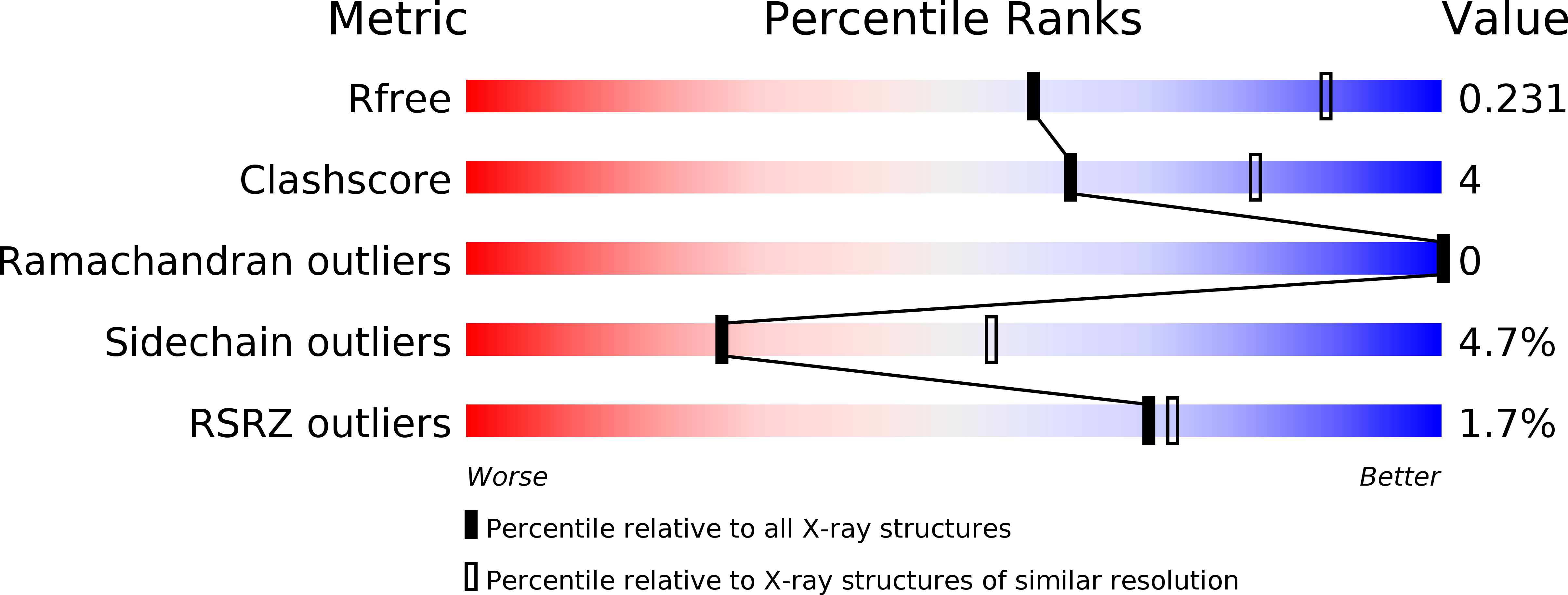
Resolution:
2.70 Å
R-Value Free:
0.22
R-Value Work:
0.17
R-Value Observed:
0.18
Space Group:
P 43 21 2
Macromolecular Entities
Structures with similar UniProt ID
Protein Blast
Polymer Type:polypeptide(L)
Molecule:50S ribosomal protein L16 arginine hydroxylase
Gene (Uniprot):roxA
Mutagens:E146A, K147A
Chain IDs:A
Chain Length:384
Number of Molecules:1
Biological Source:Escherichia coli
Modified Residue
Compound ID Chain ID Parent Comp ID Details 2D Image
MSE A MET SELENOMETHIONINE
Primary Citation
Structure and Functional Analysis of YcfD, a Novel 2-Oxoglutarate/Fe(2+)-Dependent Oxygenase Involved in Translational Regulation in Escherichia coli.
J.Mol.Biol. 426 1898 1910 (2014)
PMID: 24530688 DOI: 10.1016/j.jmb.2014.02.008

Abstact

The 2-oxoglutarate (2OG)/Fe²⁺-dependent oxygenases (2OG oxygenases) are a large family of proteins that share a similar overall three-dimensional structure and catalyze a diverse array of oxidation reactions. The Jumonji C (JmjC)-domain-containing proteins represent an important subclass of the 2OG oxygenase family that typically catalyze protein hydroxylation; however, recently, other reactions have been identified, such as tRNA modification. The Escherichia coli gene, ycfD, was predicted to be a JmjC-domain-containing protein of unknown function based on primary sequence. Recently, YcfD was determined to act as a ribosomal oxygenase, hydroxylating an arginine residue on the 50S ribosomal protein L-16 (RL-16). We have determined the crystal structure of YcfD at 2.7 Å resolution, revealing that YcfD is structurally similar to known JmjC proteins and possesses the characteristic double-stranded β-helix fold or cupin domain. Separate from the cupin domain, an additional globular module termed α-helical arm mediates dimerization of YcfD. We further have shown that 2OG binds to YcfD using isothermal titration calorimetry and identified key binding residues using mutagenesis that, together with the iron location and structural similarity with other cupin family members, allowed identification of the active site. Structural homology to ribosomal assembly proteins combined with GST (glutathione S-transferase)-YcfD pull-down of a ribosomal protein and docking of RL-16 to the YcfD active site support the role of YcfD in regulation of bacterial ribosome assembly. Furthermore, overexpression of YcfD is shown to inhibit cell growth signifying a toxic effect on ribosome assembly.

Legend

Protein

Chemical

Disease

Primary Citation of related structures